Laboratory #2: Parasitic protistans: Amebas and others
Most if not all of the animal species harbor one or more species of protozoan in their digestive tract. Although most may be considered commensals, several are parasites, and can cause considerable harm to the host. The human digestive tract may be infected with up to 12 species of protozoans. Today we will mostly focus on protozoans of the digestive tract. However, in the following three labs you will see that humans and other animals may be infected with protozoans in a diversity of other sites, including the reproductive tract, circulatory system, the skin, the brain, and elsewhere.
Protozoans of the digestive tract usually exhibit two forms, an active feeding, dividing form called the trophozoite, which does not survive for long outside of the host, and a non-feeding encysted form that can survive in the external environment. This stage is typically called the cyst.
The objective today is to study the specimens (with the aid of your textbook and other figures) and to learn the distinguishing features of each species. You will need to measure the size of each specimen you examine. Although you are required to make plates of a subset of the specimens you examine, you are encouraged to make sketches for your entries. Sketches are valuable for future reference. Be sure to make a notebook entry for each of the specimens you see.
I. Phylum Archamoebae (Amoebae) [called Phylum Rhizopoda in some editions of your text] Members of the phylum Archameobae (= Rhizopoda=Sarcodina) are characterized by their use of protoplasmic or cytoplasmic streaming to form pseudopodia that are used in locomotion. Some species in this phylum possess flagella at certain temporary life cycle stages. Most species in this phylum are free-living, but some species are parasitic and are fairly important.
Study the specimens that have been provided and note characteristics that can serve to distinguish the species in both the trophozoite and cyst stages. You should be familiar with the following features of amoebae: endoplasm, ectoplasm, nucleus, endosome (=nucleolus, but use the former term), chromatoid bars, cysta wall, food vacuole, glycogen vacuole, and pseudopod. Use the chart provided in attached Figure 1 to assist you in distinguish the various species of amoebae.
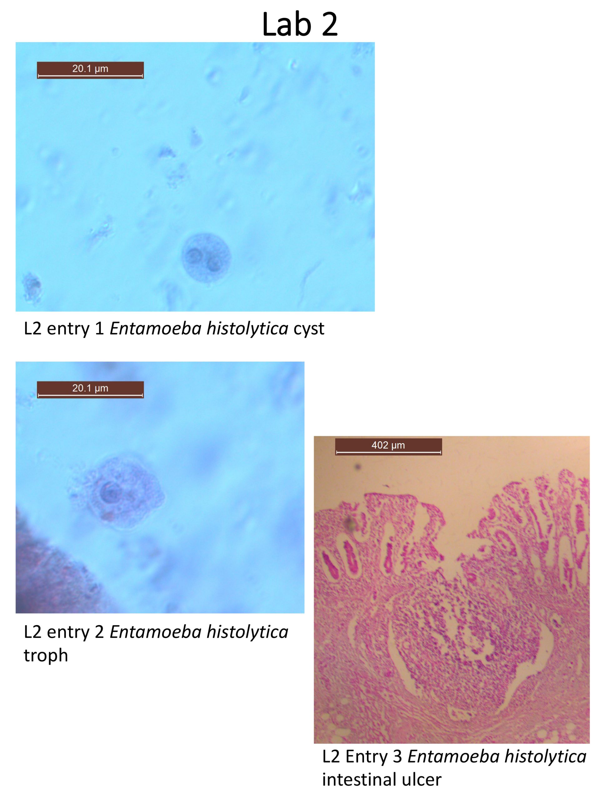
1. Entamoeba histolytica. Cyst. Fecal smear. Slide box slide 2 (See figure and textbook pg. 106 Figs. 7.1 & 7.2). You viewed this specimen in Lab 1. Locate: up to 4 nuclei, nuclear membrane chromatin, endosome, chromatoid bar, cyst wall. Note that this species possesses four nuclei and chromatoid bars that are smooth.
2. Entamoeba histolytica. Trophozoite. Fecal smear. Slide box slide 3 (See figure & text pg. 106 Fig. 7.1) You viewed this specimen in Lab 1. Trophozoites of this species can invade mucosal tissue and cause the charaxteristic flask-shaped ulcers in the wall of the large intestine (see #3 below). Red blood cells can often be observed inside the food vacuoles. Note the following structures: nucleus, nuclear membrane chromatin, endosome, food vacuole, cell membrane, and pseudopod. Note the central endosome within the nucleus and the relatively even arrangement of the nuclear membrane chromatin. These features help distinguish this potentially pathogenic species from the commensalistic Entamoeba coli.
Provide an example of a body organ other than the large intestine where you might find trophozoites of E. histolytica: ____________________________
3. Entamoeba histolytica. Section of large intestine with trophozoites. Demonstration. (text pg. 109 Fig. 7.5) This is an example of the characteristic flask-shaped ulcer. For your notebook entry, record the diameter of this ulcer, in micrometers.
What is the ontogenetic stage of the E. histolytica cells that are inside this ulcer?__________________
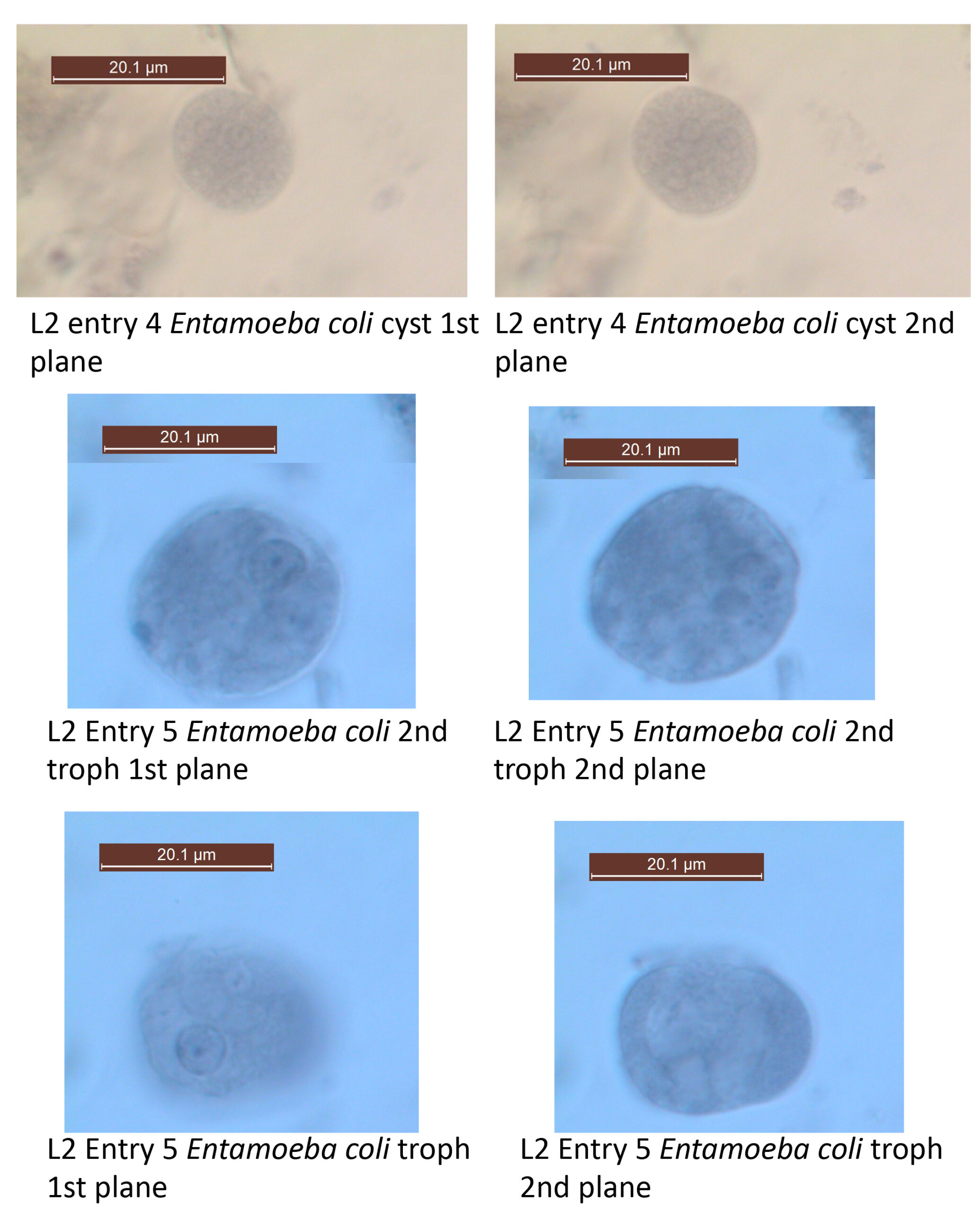
*4. Entamoeba coli. Cyst. Fecal smear. Slide box slide 4 (see figure & text pg. 112 Fig. 7.8). You studied and drew this specimen last week.
How would you distinguish cysts of this species from those of E. histolytica? Provide 2 distinguishing features:____________________________ & __________________________ .
*5. Entamoeba coli. Trophozoite. Fecal smear. Slide box slide 5 (see figure & text pg. 112 Fig. 7.7) Note how “junky” the cytoplasm is. This species often engulfs intestinal debris. Be sure to locate the nucleus, nuclear membrane chromatin, endosome, pseudopod, food vacuole & cell membrane. Note the eccentric endosome within the nucleus and the uneven nuclear membrane chromatin. Compare these features to the features of trophs of E. histolytica.
Provide an example of a potential food item of this ontogenetic stage of this organism:_______________________________
6. Entamoeba gingivalis. Trophozoite. Smear from oral swab. Demonstration. (see figure & text pg. 112 Fig. 7.9).
What is the site of infection of this species? ____________________________
How is this species transmitted? ____________________________
Does this species encyst? ____________________________
7. Iodamoeba buetschlii. Cyst. Fecal smear. Slide tray. (see figure & text pg. 113 Fig. 7.11). Note the large glycogen vacuole, large endosome and achromatic strands extending from the endosome to the nuclear membrane in this species.
What domestic animal also hosts this species? ____________________________
II. Phylum Heterolobosea
The phylogenetic relationships of many of the major prototist groupings (e.g., phyla) are still being resolved. As a result, the taxonomy (names) is changing as well. Although still called “amebas” and treated with other ameba species in your textbook, the following two species belong to a different grouping known as Heterolobosea.
8. Naegleria fowleri. Trophozoites in brain tissue. Histological section. Demonstration.
(text pg. 115 Fig. 7.13 & 7.14). This is an example of a facultative parasite. This species normally lives in soil and water but can facultatively (i.e., if given the chance) become parasitic. As a parasite it produces primary amoebic meningoencephalitis (PAM) in humans and can result in death. This amoeba is unusual in its ability to produce a cyst and a biflagellated form, in addition to the amoeboid trophozoite.
What is the normal habitat of these organisms?________________________
III. Phylum Retortamonada
The members of this phylum are distinctive in that they lack mitochondria and golgi. Most species of this phylum are inhabitants of the intestinal tracts of vertebrate hosts. All species in this group, whether parasitic or free-living, inhabit anoxic environments. Most species possess a resistant cyst stage and a more fragile feeding stage called the trophozoite.
Unfortunately, we have only a single species of the phylum to show you in lab today. But be sure to keep in mind there are several other species that can be of veterinary or commercial significance.
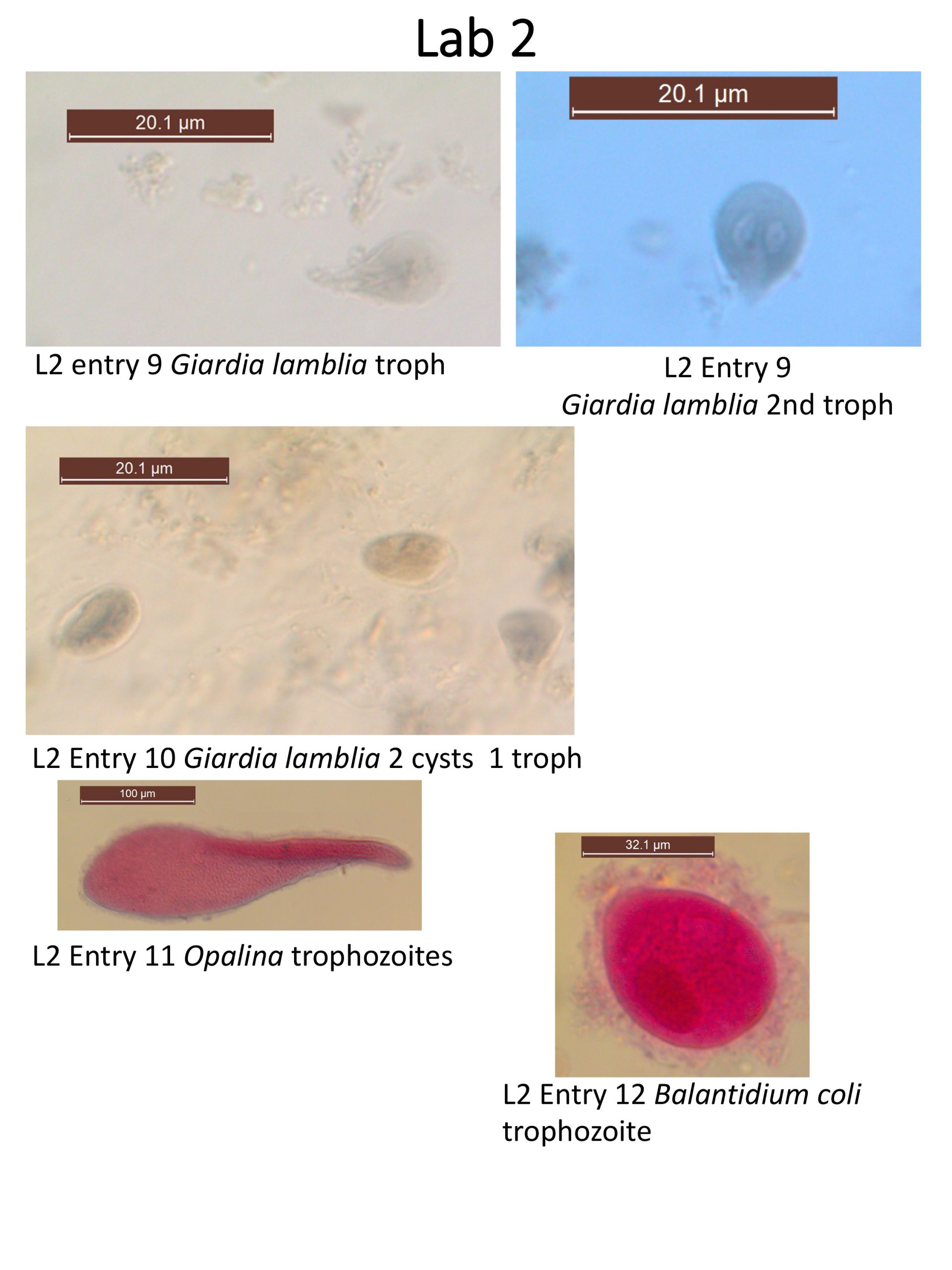
9. Giardia lamblia / Giardia duodenalis Trophozoite. Fecal smear. Slide box slide 6 (see figure & text pg. 89). This species can be pathogenic in humans. This species and its close relatives can also be found in the intestinal tracts of a diversity of other mammal species. Recognize the following structures: nuclei, adhesive disc, median bodies (if visible), anterior flagella, posterior flagella, ventral flagella, and caudal flagella.
Where , in what body organ, would you expect to find this stage of this species? ______________________
What structure does this organism remain attached to its host? ________________________
10. Giardia lamblia / Giardia duodenalis . Cyst. Fecal smear. Slide tray. (see figure & text pg. 89). Locate the following: nuclei, cyst wall, flagella, and median bodies (if visible).
Where would you expect to find this stage of this species? ________________________
V. Phylum Chromista
The opalinids possess numerous rows of flagella over the entire body surface and consequently have traditionally been placed with the Ciliophora. However, recent detailed studies with electron microscopy suggest that they are different from ciliophorans in several ways. For example, they lack infraciliature and possess two or many nuclei of similar structure and size. There is some evidence that suggests reproduction of opalinids is controlled or cued by host hormones.
11. Opalina. Trophozoites. Whole mount from frog rectum. Slide tray. (see figure & text pg. 102 Fig. 6.18). Note the numerous nuclei of similar size and shape as well as the many flagella.
How does this species get transmitted from host to host? That is, how does it leave the body of one host, and how does it enter the body of the next host? _______________
VI. Phylum Ciliata – (ciliates)
Ciliates possess multiple cilia that are united via a network of organelles known as infraciliature, which lies below the pellicle. They also possess two types of nuclei (usually a macronucleus and a micronucleus). A contractile vacuole is usually also present.
12. Balantidium coli. Trophozoites. Fecal smear. Demonstration. (see figure & text pg. 168 Figs. 10.2 & 10.3). This is the largest protozoan parasite of humans. Note the sausage-shaped macronucleus, round micronucleus, and numerous cilia.
What structures would this organism use for locomotion? ______________________