Laboratory #8: Platyhelminthes II (lung fluke, blood flukes, and flukes of wildlife)
Phylum Platyhelminthes — flatworms
Subphylum Neodermata
Class Trematoda
Subclass Aspidogastrea
Subclass Digenea
Last time you were introduced to digenean life cycle stages and the general morphology of an adult digene, and you also observed liver flukes. Now you will become familiar with lung flukes and blood flukes, which are some of the most detrimental parasites known. Bear in mind that many of the species of this highly diverse group are parasites of other animals. Also keep in mind that nearly every species we discuss utilizes a snail as the first intermediate host.
Lung fluke
The lung flukes are usually found associated with the lungs of their definitive hosts. Our lung fluke example is Paragonimus westermani. Lung flukes can be found in a variety of hosts, such as a species in bullfrogs that they acquire by consuming dragonflies.
1. Paragonimus westermani eggs. Demonstration. Fecal smear. (see text pg. 271). Note the distinctive shape of this egg as well as the operculum and characteristic “shoulders” at the junction of the operculum with the rest of the shell.
How large is this egg?____________________________
Recognize these structures: operculum, shell, and shoulders.
How do eggs of this species leave the body of the definitive host? __________________
How would you distinguish eggs of this species from those of C. sinensis or F. hepatica?_______________________________________
2. Paragonimus westermani in lung tissue section. Demonstration. (see life cycle on text pg. 270). Worms inside the lung stimulate an inflammatory response that ultimately results in the worms becoming enshrouded in a capsule of scar tissue. Study the section for eggs and worm fragments of P. westermani
Will eggs produced earlier or later be more likely to be transmitted to the next host in the life cycle? Explain:_________________________________________________
3. Paragonimus westermani adult. Demonstration. Whole mount. (see text pg. 269). Lung parasite is the etiological agent of paragonimiasis in humans. This is a good example of a zoonosis; P. westermani typically parasitizes wild animals—especially cats—but causes disease when it infects humans. Observe the lobed testes and ovary, which is slightly off the center of the body. There is a short uterus and suckers are inconspicuous. The overall size and shape of this specimen is similar to a coffee bean.
What are the intermediate hosts in the life cycle of Paragonimus westermani?__________________________________________________
Blood flukes
Family Schistosomatidae
These kinds of flukes parasitize the circulatory system of their definitive host. The schistosomatids parasitize warm-blooded definitive hosts while other families of blood flukes parasitize cold-blooded definitive hosts, such as fish, elasmobranchs, or reptiles.
All species in this family are unusual for digeneans in that they are dioecious. In addition, members of this family are unusual in that they lack a metacercarial stage in their life cycle, rather, cercaria locate, and subsequently penetrate, the definitive host directly. Three species in this family are notoriously important parasites of humans. Recent work suggests that several other species may also regularly parasitize humans, but their geographic distribution and prevalence is much lower than the 3 species we will view.
Many other species in this group parasitize birds or other groups of mammals. These schistosome species are responsible for a condition in humans known as “swimmer’s itch” or “clam digger’s itch,” when cercaria of these species attempt to penetrate the skin of unsuitable (human) hosts.
Human schistosomes
Schistosoma mansoni adults Text pages 238-249 for full coverage; for today focus on the first three pages. Examine separate slides of males and females, as well as a slide of the 2 sexes together en copula. Make a plate of the worms en copula.
4A. Adult male. Demonstration. Locate oral sucker and acetabulum (ventral sucker).
Do you see an intestine?_________________________________
4B. Adult female. Demonstration. Compare the female to the male and note the difference in morphology between the 2 sexes.
Which sex is more robust? __________________________
Which is slighter?________________________________
*4C. En copula. Slide box slide #24. Note the oral suckers and acetabulum of both individuals. Also note the female’s position in the gynecophoral canal. Label any other structures you can recognize, and take special care to label which worm is male and which is female.
Can you see a feature of the male’s outer body that would enable it to maintain its position in its habitat? ______________________________________________
If so, what is the feature? __________________________________________
Does the female also have this feature?___________________________________
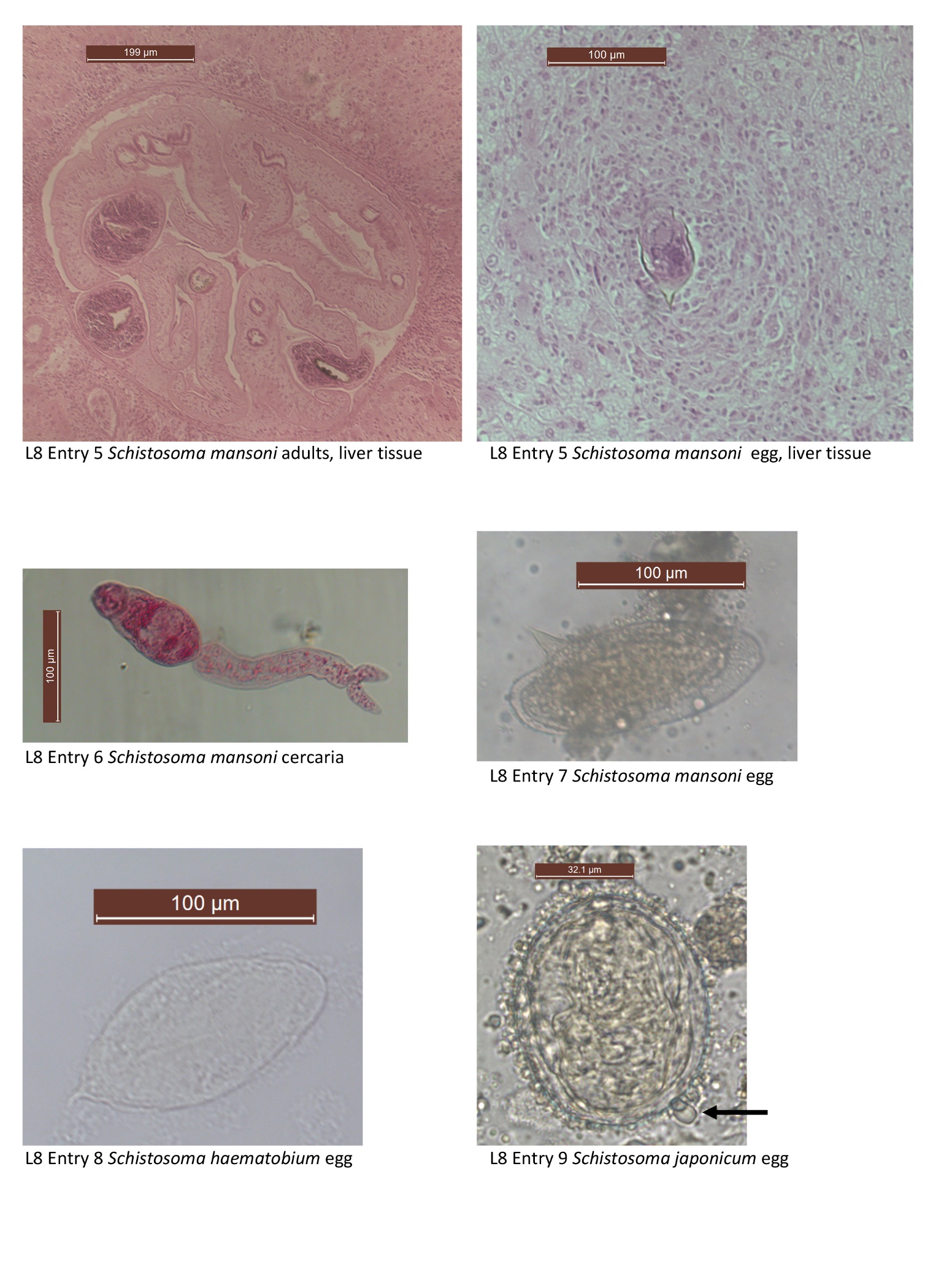
5. Schistosoma mansoni adults and eggs in section of liver tissue. Demonstration. (see text pg. 246 fig. 16.16). It is important to keep in mind that there are two sources pathology associated with schistosomiasis, adult worms, and the eggs they produce. Look for evidence of pseudotubercles, a type of granuloma caused by Schistosoma eggs.
How can you recognize these eggs as Schistosoma mansoni? You may wish to wait until after completing entry #7 to answer this._______________________________________
6. Schistosoma mansoni cercaria. Slide box slide 20. (see text pg. 241). You examined these last week. This is a typical schistosome cercaria: it lacks a pharynx, and has a forked tail.
How would this specimen locate the next host in its life cycle?__________________
Look on page 242 for images of the following 3 egg species:
*7. Schistosoma mansoni eggs. Fecal smear. Slide tray. (see text pg. 242). Note the characteristic lateral spine on this egg.
What is the size of the egg?_______________________________
Make a drawing and label: miracidium (if one is present and visible), shell, and lateral spine.
Do you see evidence of an operculum?________________________________
How do miracidia exit these eggs? (Hint, see fig. in text pg. 243)__________________
By what means do these eggs exit the definitive host?_________________________
*8. Schistosoma haematobium eggs. Urine smear. Slide tray. (see text pg. 242). Note the characteristic terminal spine on this egg.
What is the size of the egg?_______________________________
Do you see evidence of a miracidium in any of these eggs?__________________
Make a drawing and label: miracidium (if one is present and visible), shell, and terminal spine.
By what means do these eggs of this species exit the definitive host?_________________
How does this egg differ from an egg of S. mansoni?_____________________________
*9. Schistosoma japonicum eggs. Fecal smear. Slide tray. (see text pg. 242). Note the characteristic rudimentary lateral spine. It is less prominent than the spine of S. mansoni. on this egg.
What is the size of the egg?__________________
Make a drawing and label: miracidium (if one is present and visible), shell, and lateral spine.
How does this egg differ from the previous 2 species?____________________________
How do eggs of this species exit the definitive host?_____________________________
Flukes of other animals
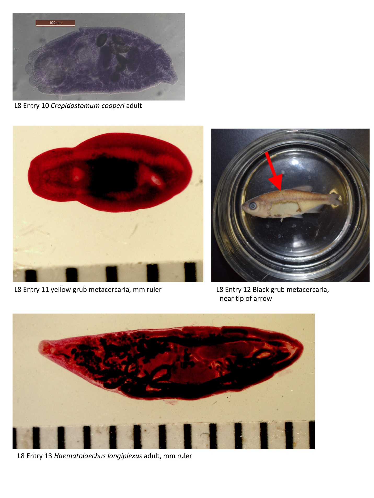
10. Crepidostomum cooperi from Largemouth bass intestine. Demonstration. This digenean species was collected from the Thayer Farm Big Pond at the Biological Field Station—the location of our field trip later this semester. The first intermediate host in the life cycle is C. cooperi is a fingernail clam. Cercaria emerge from clams until they find mayfly larvae (also aquatic). The life cycle is completed when a fish consumes an infected mayfly, which serves as the definitive host. Look at the specimen on demonstration. This species is characterized by numerous papillae surrounding the oral sucker.
What is different about the hosts in the life cycle of C. cooperi, compared to other digeneans we have discussed?_________________________
11. Clinostomum marginatum (or “Yellow grub”) metacercaria from muscle of a Pumpkinseed sunfish. Demonstration. This is a metacercaria that is commonly found in local fish of economic importance (e.g., Largemouth bass). Metacercaria of C. marginatum do not encyst, rather, they can be found freely creeping about the body cavity of the fish, or in the muscle, or on the fins. Fish are considered the second intermediate host. Herons serve as the definitive hosts, while certain snail species serve as the first intermediate host.
How might metacercaria differ from adult specimens of C. marginatum?_________________________________________________________
What do you suppose would happen if you consumed metacercaria (e.g., imagine having largemouth bass sushi)?__________________________________________________
Can this parasite “travel” from lake to lake? How?_____________________________
12. “Black grub” on external surface of a minnow. The visible black spots are not actually grubs; they are dead metacercaria. We cannot know the species identity of these parasites because they are immature worms and because they are largely destroyed by the host immune response. That is, there are a variety of trematode species that could be responsible for “black grub” in fish.
What is the term for the host role that this fish plays in the life cycle of the “black grubs” visible here?___________________________
13. Haematoloechus longiplexus from lung of a bullfrog. Demonstration. (see similar species in Fig. 18.4 in text pg. 268). This adult specimen was collected from a bullfrog lung, in Nebraska. Haematoloechus species are lung flukes, i.e., they live and mate as adults in the lung tissue of frogs. First and second intermediate hosts in the life cycle are snails and dragonflies, respectively. Dragonflies acquire cercaria via jet-like propulsion into their anal gills. Frogs acquire worms when preying on dragonflies. We have collected specimens of Haematoloechus from all three hosts in the life cycle, at a tiny mucky pond at the Biological Field Station.
What might be the food item of adult H. longiplexus?_________________________