Laboratory #12: Miscellaneous parasitic phyla
Phylum Acanthocephala (now in Phylum Rotifera)
Acanthocephalans, or “thorny-headed worms”, inhabit the intestines of fishes, mammals, birds, and rarely amphibians and reptiles, and use their hooked proboscis to attach to the host intestinal wall. The acanthocephalans resemble nematodes in their possession of a fluid-filled body cavity referred to as a pseudocoelom (or blastocoelom). Acanthocephalans lack a digestive system, as was the case with cestodes. The muscular, nervous and excretory systems of acanthocephalans are poorly developed. Acanthocephalans are called thorny-headed worms because of their most prominent feature, a thorny eversible proboscis.
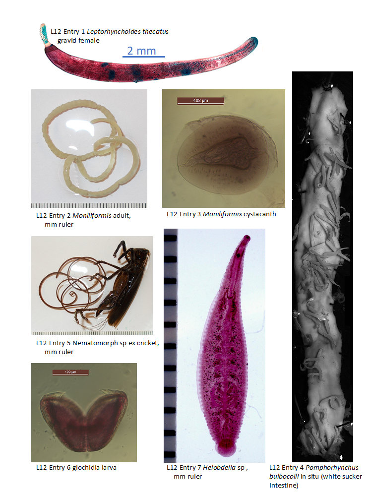
*1. Leptorhynchoides thecatus adult worm ex Largemouth bass. Slide box slide #1. View the anatomy of your specimen in comparison to the one depicted in the anatomical diagram in your textbook (See page 476). Note the armed anterior proboscis with its well-developed muscles attached to the posterior end of the proboscis sheath. The lemnisci, which appear as sac-like structures extending into the blastocoel, are thought to serve a hydrostatic function in the protrusion and retraction of the proboscis. For your drawing, only draw the proboscis, which should be viewed at 400x magnification. In addition, provide a detailed drawing of one hook, at oil immersion. You need to decide whether your specimen is a male or female, and then be sure to look at an example of the other sex from one of your lab partners. Females: Observe the female egg masses, if present, floating free in the blastocoel where fertilization and early development has taken place. Eggs pass though a funnel-like uterine bell to the uterus and vagina. The genital pore is terminal. Males: Recognize the 2 rounded testes at the anterior end of the ligament. Posterior to the testes are 8 cement glands. These secrete a substance that prevents the female from accepting sperm from other males post-copulation. There is an eversible copulatory bursa, and also a penis, which is used to transfer sperm to the female. Be sure to recognize key structures, and to determine whether your specimen is male or female.
How does this organism acquire its nutrients?___________What other kind of parasite acquires its nutrients in a similar way?____________
2. Moniliformis moniliformis adult specimen ex rat. Demonstration. This specimen was collected from the small intestine of a Norway rat in a wharf just a few blocks from the French Quarter of New Orleans. Can you see the anterior proboscis? It is small. Using the highest magnification available on the dissecting microscope, observe the individual hooks on the proboscis. This rat was infected with Moniliformis by consuming a cockroach that was infected with a larval stage known as a cystacanth, shown below. Once the cystacanth entered the rat, it grew into the female adult worm shown here. If a male was available, there would be mating and later the female would pass her eggs which would leave the rat via its feces.
What did the rat do to become infected with this parasite? _____________________________
3. Moniliformis moniliformis cystacanth stage. Demonstration. This specimen was collected from the body cavity of a cockroach. It has a proboscis that is invaginated. Once transmitted to rats, this stage grows into an adult worm. This stage of this parasite is known to change the behavior of its cockroach host, making it more likely that a rat would be able to capture a cockroach and eat it.
How do you suppose cockroaches become infected with Moniliformis? ________________________________________
4. Pomphorhynchus bulbocolli adults attached to intestine of white sucker fish. Demonstration. This acanthocephalan species can be found locally, in white suckers. White suckers are fish with ventrally-oriented mouths that feed on a variety of invertebrates in the bottom layer, including amphipods, the intermediate host of P. bulbocolli. Pomphorhynchus bulbocolli is unusual among acanthocephalans in that it pokes its proboscis entirely through the intestinal wall. This can result in extensive pathology (damage) to the fish intestine–a topic that was the focus of one research project in my lab (see included publication on display).
Provide an estimate of the number of parasites visible at this demonstration:____________
Phylum Nematomorpha
The nematomorphs, or “horsehair worms” are fairly common in freshwater. A few species are marine. This parasitic phylum is unusual in that the adult stage is actually free-living but the larvae are parasitic in the haemocoel of insects that are associated with water. Free-living adult worms mate and produce large quantities of eggs that grow into larvae that find and penetrate the appropriate host. Once in the host, larvae feed until ready to exit the host, which is done when the host goes near water.
5. Unidentified adult nematomorph emerging from grasshopper. Demonstration. See text page 465 Figs. 31.1a-b. Note the resemblance of this specimen to nematodes. They can be distinguished from nematodes on the base of their thick, often brown cuticle, and especially by lack of a mouth and possession of a bluntly rounded anterior end.
What stage in the life cycle stage of nematomorphs is parasitic?__________________
Phylum Mollusca
Molluscs are a hugely diverse phylum (~90,000 species; 2nd only to arthropods) that includes clams, snails, slugs, chitons, tusk shells, squid, octopuses, cuttlefish and nautiluses, among others. Most mollusks are free-living species, but there are several examples of parasitism within the phylum. Today we will focus on members of the freshwater clam family Unionidae. Unionids are free-living as adults, but possess a larval stage called the glochidia larva, that is an obligate parasite of freshwater fishes.
*6. Glochidia larvae. Slide tray. This larval type is restricted to members of the freshwater clams belonging to either the unionid or margaritiferid families. Note the byssal thread if visible (increase contrast on your microscope), and hooks that are used to attach to the freshwater fish host. Glochidia larvae tend to be fairly host specific. These clams cannot develop in the absence of fish because the glochidia are obligate parasites on the gills and skin of fish.
What structures are visible that would not be present in the adult stage of this animal?_____________________________
Which stage in the life cycle of the freshwater mussels travels/disperses a greater distance, the adult (free-living) stage, or the glochidia (parasitic) stage?____________________________
Phylum Annelida
Annelids include earthworms, other segmented worms (including many marine forms, e.g., the feather duster worm in Dr. Lentz’s tank), and the leeches. The most common parasitic annelids are members of the Hirudinea group, also known as the leeches. Leeches are monoecious.
7. Helobdella sp– adult. Demonstration. See attached Figure 3 for an illustration of a similar species. Although many are free-living scavengers, many are blood-feeding parasites. For example, in our fish-parasite research we frequently see leeches attached to yellow perch. The leech shown here is actually considered an opportunistic feeder rather than obligately parasitic. It can feed on other leeches for their blood meal, or on soft tissues and body fluids of other invertebrates such as insect larvae, snails, aquatic oligochaetes, crustaceans and bivalves. It also uses open wounds of vertebrates to feed on the lymph.
Note the general features of this specimen by comparing it to Figure 3. Recognize the posterior sucker, mouth, and extensive cecum.
How would you distinguish a leech from a monogene?__________________________