Laboratory #10: Nematoda I
Phylum Nematoda
Many nematodes occur as free-living animals in the sea, freshwater, and soil, but there are thousands of species that are parasitic in plants and animals. Human diseases caused by nematodes include Onchocerciasis, Filariasis, Dracunculiasis, Ascariasis and hookworm disease. These are more common in tropical countries, especially in overpopulated areas where sanitation is a problem. But, even in countries like the United States, pinworms flourish among children of rich and poor alike, and Trichinosis (acquired by eating undercooked pork) is not uncommon. Nematodes also cause numerous plant diseases (e.g. root knot nematodes), and others serve as vectors for plant viruses. Our focus here is on nematodes of vertebrates, especially those species that infect humans.
Most nematodes are dioecious. Most species possess four juvenile stages prior to the adult stage. These five life cycle stages are separated by molts. Nematodes possess a fluid-filled body cavity known as a blastocoel (or pseudocoel). Male nematodes have one or two spicules, which aid in copulation. Nematodes possess a thick cuticle and move in a serpentine fashion. Nematodes lack a circulatory system, and the excretory system may consist of a pair of specialized cells, or in the case of Ascaris, consist of a pair of lateral excretory canals. Nematodes possess a prominent digestive system, and are said to have a “tube within a tube” body plan. The digestive system includes a long straight intestine, a rectum, and a subterminal anus. Thus the gut is considered complete.
The two nematode classes are distinguished on the basis of sensory organs, which are difficult to see without the aid of an electron microscope. Both classes include free-living and parasitic species:
- Class Chromadorea / Order Rhabditea (=Secernentia =Phasmidea) – possess cephalic amphids and caudal phasmids
- Class Enoplea (=Adenophorea) – only possess cephalic amphids
I. Class Chromadorea / Order Rhabditea (=Secernentia =Phasmidea)
Most species in this group are soil-dwelling forms. It seems likely that the parasitic species in this group evolved from free-living ancestors.
Infraorder Ascaridomorpha
These kinds of nematodes are typically large, stout, intestinal parasites with three lips, but there are a few exceptions. The esophagus is usually muscular, and simple (without a conspicuous bulb). The life cycle is usually monoxenous.
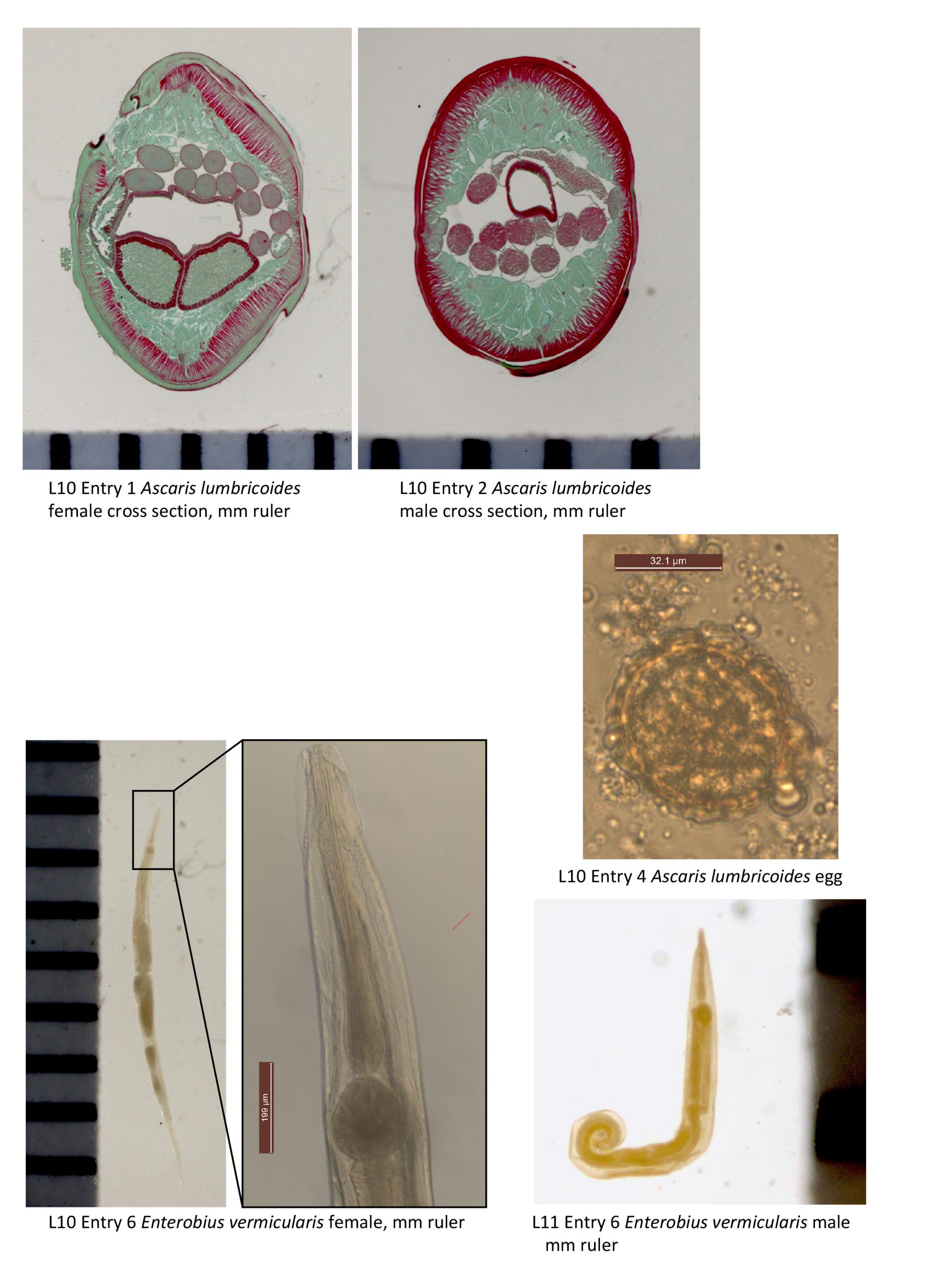
Cross sections as examples of general nematode structure:
*1. Ascaris lumbricoides. Cross section of adult female. Slide box slide #28. See text page 355. Fig. 22.11b. These cross sections illustrate some of the key features of nematodes. Make a full-page plate of a section through an adult female A. lumbricoides. The outer body layer is the cuticle, which surrounds the epidermis. Inside the epidermis is the hypodermis, the layer that contains the lateral excretory canals. Muscle cells are seated on the hypodermis and have a striated contractile portion and a non-contractile portion that has the nucleus and processes that extend to the dorsal or ventral nerve cords. Neuromuscular junctions of nematodes are unique; rather than possessing nerves that run to muscles, as in most other animals, the muscles of nematodes possess non-contractile portions that extend to either the dorsal or ventral nerve cord.
Locate the following structures on your plate: cuticle, hypodermis, lateral excretory canals, dorsal nerve cord, ventral nerve cord, uterus, egg, ovary, oviduct, blastocoel, intestine, and muscle cells.
What features are useful for distinguishing between sexes:_______________________________________________________
List 3 of the differences between this nematode and species of platyhelminths:_____________________________________________________
Because the reproductive tract is coiled, you may see several sections of the uterus, ovaries and the oviducts (in the section of a female) or portions of the testes and vas deferens (in the section of the male).
2. Cross section of adult male. Slide box slide #28. See text page 355, Fig. 22.11a. Compare the cross section of the male worm to the female worm that you just drew. Note that the testes and vas deferens are present instead of the female reproductive structures. All the remaining structures should be familiar to you based on the examination of the female.
3. Ascaris lumbricoides. Demonstration of adult male and female worm. See text page 412, Fig. 26.1. Note the size of these worms. If a dissected male is available, recognize: Testes, intestine, lateral lines. In females, recognize: Intestine, vagina, uterine branches, oviducts, ovaries, and lateral lines. Be able to distinguish males from females based on external appearance.
How could you distinguish the sexes of this species from an external view?_____________________________________________________________
Note the lateral lines (=lateral epidermal cord) running the length of individuals of both sexes. What structures do lateral lines contain?_________________________________
Look for evidence of muscle cells. Also locate the dorsal and ventral nerve cords.
*4. Ascaris lumbricoides. Eggs. Slide box slide #29. See text page 412 Fig. 26.2 & 26.3. Note the rough, mammillated layer laid down by the uterus. Make a drawing of an egg and label the following: developing embryo and mammillated layer.
How do fertilized and unfertilized eggs differ? ___________________Refer to page 412.
5. Toxocara cati. Adult worms. Demonstration. See text Fig. 26.10 for image, and Fig. 26.7 for life cycle of similar species, Toxocara canis. This specimen was collected a cat that was dissected during an Anatomy & Physiology lab in this department. The worms were collected from the stomach, but their actual location in the living cat would have been the small intestine. These worms look similar to Ascaris species, but are much smaller, and have a pair of lateral projections at the anterior end known as cervical alae. Toxocara canis and Taxocara cati are nematodes of our domestic household pets. However, these species can infect humans, an improper host, and cause a condition known as visceral larval migrans, in which juvenile worms migrate throughout tissues, causing a variety of problems.
Why do you suppose these worms were actually encountered in the cat’s stomach, rather than the small intestine?_______________________
Infraorder Oxyuridomorpha
Members of this order are commonly known as “pinworms” because the females, in particular, have sharp, pointed tails. Males in this group possess a single spicule. Male pinworms are haploid and females are diploid, this condition is known as haplodiploidy. Pinworms are further characterized in having a prominent muscular bulb at the posterior end of the esophagus. Life cycles of oxyurids are usually direct, i.e., single-host.
6. Enterobius vermicularis adult female. Demonstration. See text page 446 Fig. 27.1 and 27.4. Locate the anterior lips (3), cervical alae (anterior cuticular inflations) esophageal bulb, pseudocoelom, uterus, vagina, egg, cuticle, and anus.
How are the eggs of this species transmitted?_________________________________
7. Enterobius vermicularis adult male. Demonstration. See text page 427 Fig. 27.3. Note the mouth surrounded by 3 lips, anterior cuticular inflations (cervical alae), and the presence of a spicule. Identify the structures in attached Figure 3.
How would you distinguish between individuals of the two sexes of this species?
_______________________________________________________________
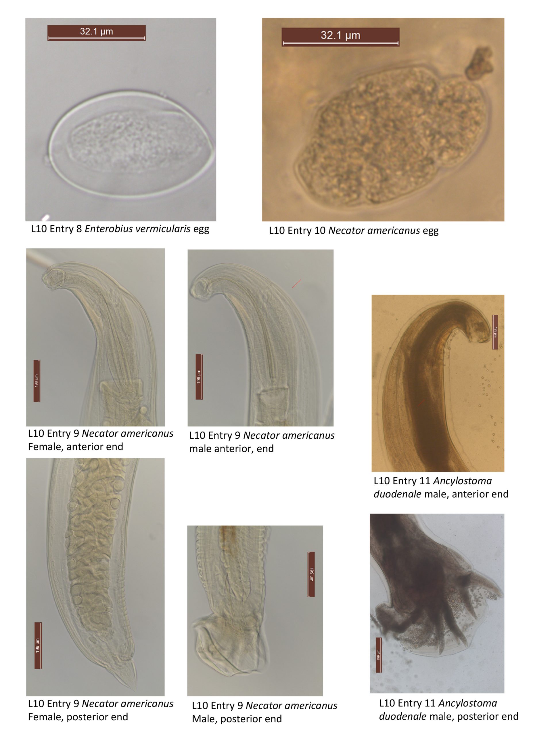
8. Enterobius vermicularis eggs. Demonstration. See text page 427 Fig. 27.5. Eggs of this species can be recognized in that they are flattened on one side. Sketch this egg in your notes and label the shell, and developing JI inside the shell.
Infraorder Rhabditomorpha
Male individuals belonging to this group generally have a broad copulatory bursa (i.e., a bag) supported by a number of rays. See Text 418 Fig. 25.2 to become familiar with the morphology of a copulatory bursa.
Most species of this order are intestinal parasites of vertebrates as adults, and are monoxenous.
Family Ancylostomidae
Members of this group are known as the “hookworms”. They typically have a sclerotized buccal capsule that is armed with cutting plates or teeth. They attach to the intestinal mucosa with this capsule and feed on blood and tissue fluids sucked from the mucosa.
*9. Necator americanus– Adult male and female. Slide box slide #30. Text page 400 Figs. 25.6 & 25.7. This species parasitizes humans. It is characterized by the pairs of dorsal and ventral cutting plates at the anterior margin of the buccal capsule. It also has a pair of subdorsal and subventral teeth at the rear of the capsule. Compare the morphological differences between male and female N. americanus specimens. Note the copulatory bursa in the male; the spicules in this species are elongate and are fused distally.
10. Necator americanus– eggs. Demonstration. Text page 399 Fig. 25.4. The eggs of hookworms share the same general features. The eggs are laid containing embryos in the 2, 4, or several cell stage of development, which pass to the outside with the feces of the host. Note the egg shell and cells of a developing embryo. Make a drawing of an egg of this species. Label the egg shell and cells of the developing embryo.
On average, in what cell stage of cleavage are the embryos on your slide?_________
11. Ancylostoma duodenale– Adult male. Demonstration. Text page 401 Figs. 25.8 & 25.9. This species also parasitizes humans. It has a buccal capsule with anterior plates, each two bearing two large teeth. The spicules in male A. duodenale are elongate, but are not fused distally.
Do you see evidence of a copulatory bursa in this species?________________________
How could you distinguish this species from N. americanus?______________________