Laboratory #3: trypanosomes and their kin
Phylum Parabasalia
The members of this phylum possess a conspicuous axostyle or stout rod composed of microtubules that runs from the kinetosome to the posterior end of the trophozoite. Some members of this phylum lack mitochondria.
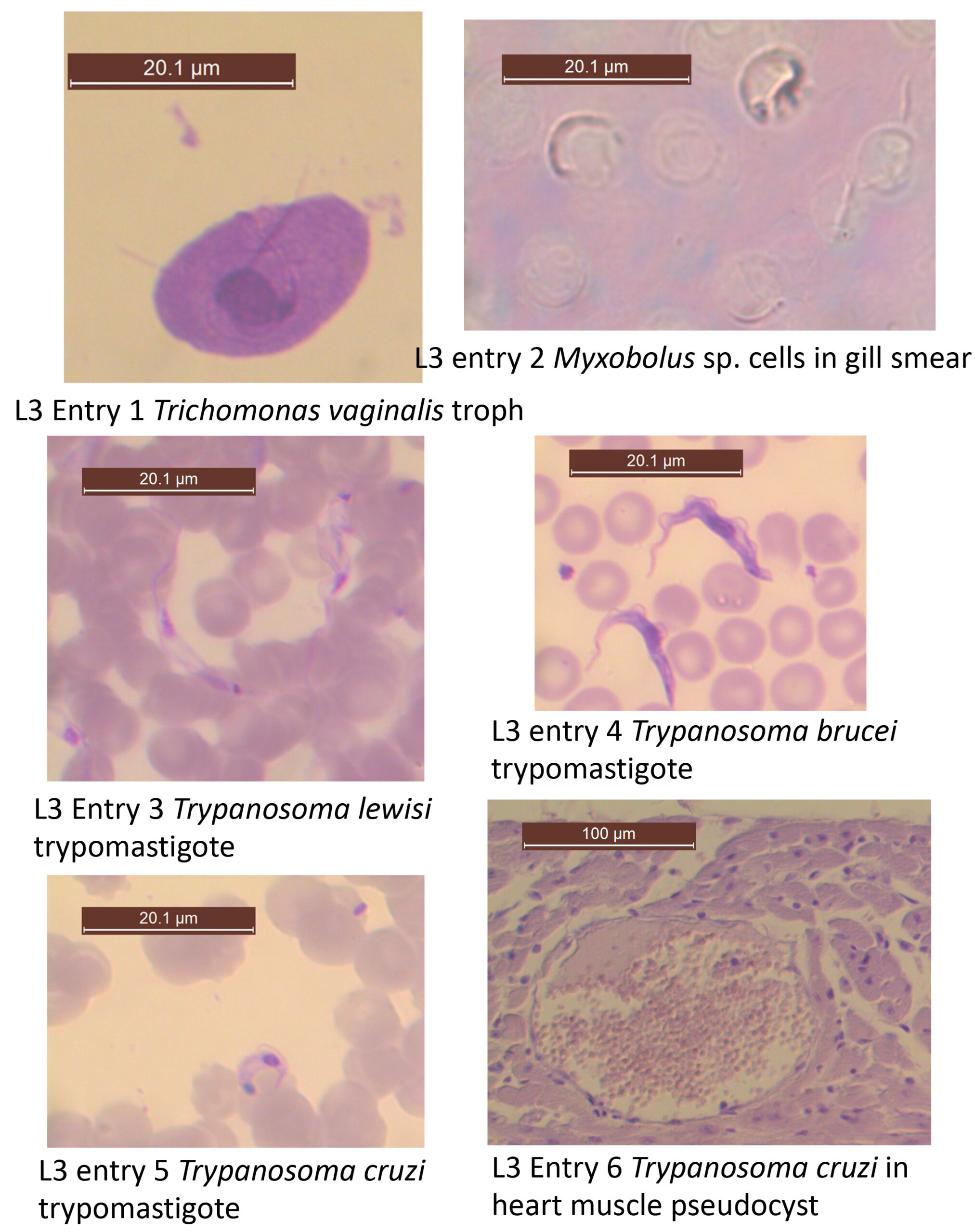
*1. Trichomonas vaginalis. Trophozoite. Vaginal smear. Slide box slide 7 (see figure & text pg. 95 Fig. 6.12). Species of Trichomonas are part of a group of the phylum Parabasalida that lack mitochondria, do not form cysts, and typically possess 4 or 6 flagella. Recognize the following structures: nucleus, kinetosome, anterior flagella, undulating membrane, recurrent flagellum (note extent), costa, and axostyle. Note: for your drawing use a cell that is not in the midst of fission!
Where would you expect to find this species in its host? _____________________
What is the function of the flagella that you can see? _______________
Phylum Cnidaria – cnidarians including myxozoans
Cnidarians make up a diverse phylum of 1000s of species that are an important part of sealife and familiar animals to you, including jellyfish, Portuguese man o’ war,
anenomes, and coral. But why are they mentioned in this lab exercise? Phylum Cnidaria is now known to include a group of single celled parasitic organisms known as the Myxozoa. Myxozoa, or myxozoans have traditionally been considered their own phylum. With the advent of the use of DNA sequence data to study patterns of evolutionary relatedness, however, it has come to light that myxozoans are actually cnidarians! In other words, the single-celled parasites known as myxozoans, are actually more closely related to jellyfish, than they are to other “protistan” parasites.
- Myxobolus sp. Gill smear. Demonstration. (text pg. 178 Fig. 11.3). This specimen was found attached to the gills of a Quilback (a type of fish in the sucker family) from western Nebraska. Examine the specimen and recognize the polar filaments. Polar filaments are basically modified cnidarian nematocysts. This thread-like structure was shot out of the cell and is used for attachment to the fish gill. The previous host in the life cycle would have been an aquatic worm (oligochaete annelid).
Consider the specific habitat of these parasites. What would be a possible pathological effect on the fish host, caused by the presence of Myxobolus?_____________________
Phylum Euglenozoa
Members of this phylum typically have one or two flagella and mitochondria with discoid (instead of tubular) cristae. This phylum includes many free living species, as well as various species that are parasites of plants or animals.
Subphylum Kinetoplasta
Members of this subphylum are very distinctive in their possession of a unique mitochondrion containing a large disc of DNA composed of both mini and maxi circles, commonly referred to as a kintoplast.
Order Trypanosomatida
The species in this group possess a single flagellum that is usually attached to the body via an undulating membrane. Most species in the group are heteroxenous; their life cycles include both an invertebrate and a vertebrate host. These are among the most pathogenic species of flagellates. You will see representatives of two trypanosomatid genera, Trypanosoma and Leishmania.
I. Trypanosomatida exhibiting a trypomastigote form
Be sure to use the accompanying figures in your textbook as you study the characteristics that help differentiate trypomastigotes of different species. Be sure to take measurements whenever possible. Remember that sketches are useful to refer to in the future.
3. Trypanosoma lewisi– Trypomastigotes. Rat blood smear. Slide tray. (text page 76 Fig. 5.15). These specimens are excellent examples of trypomastigote morphology. Locate the following: nucleus, kinetoplast, kinetosome (if visible), undulating membrane, and free flagellum. Note that the kinetoplast is posterior to the nucleus in position and the very long undulating membrane that are characteristic of this trypanosomatid body form.
*4. Trypanosoma brucei gambiense or T. b. rhodesiense or T. b. brucei– Trypomastigotes. Mammal blood smear. Slide box slide 8 (text page 63 Fig. 5.3f). Although these three subspecies are the etiological agents of very different diseases, their trypomastigotes are morphologically indistinguishable. This is why some researchers consider all 3 taxa to be subspecies of the species Trypanosoma brucei. Note: in your drawing you should include 1-2 red blood cells for context.
Typomastigotes have two forms. Which form do you see on the slide? ____________
Fill in the table below with details on the hosts of the 3 subspecies of Trypanosoma brucei:
|
Characteristic |
Trypanosoma brucei brucei |
Trypanosoma brucei rhodesiense |
Trypanosoma brucei gambiense |
|---|---|---|---|
|
Possible definitive host |
|
|
|
|
Intermediate host species (be specific) |
|
|
|
What other life cycle stage, besides the trypomastigote, is present in the life cycle of all of these subspecies?__________
*5. Trypanosoma cruzi– Trypomastigotes. Human blood smear. Slide box slide 9 (text page 71 Fig. 5.8). Trypomastigotes of this species have a characteristic “C” shape. Find the nucleus, kinetoplast, kinetosome (if visible), undulating membrane, and free flagellum. Note: in your drawing you should include 1-2 red blood cells for context.
How does the geographic distribution of this species differ from that of the 3 subspecies of T. brucei?____________________________________________________________
6. Trypanosoma cruzi– Amastigotes. Section of infected heart muscle. Demonstration (text page 73 Fig. 5.11). Concentrated pockets of amastigotes of T. cruzi are often referred to as “pseudocysts.”
What other ontogenetic stage(s), besides amastigotes, of this species is found in humans?__________________
II. Trypanosomatida lacking a trypomastigote form
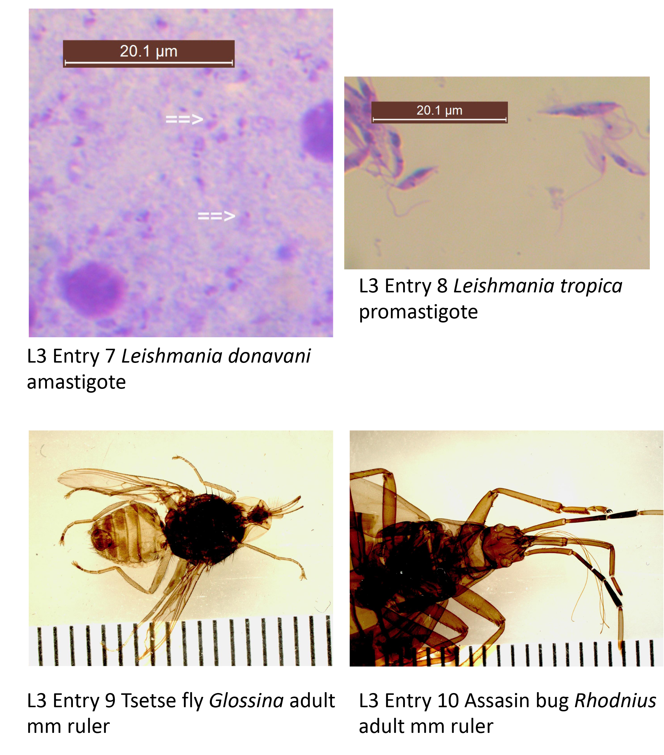
*7. Leishmania donavani– Amastigotes. Hamster spleen smear. Slide tray. (text page 78 Fig. 5.18). Note the tiny size of these organisms; they are among the smallest known eukaryotic cells. Also note the shape of the cell, the conspicuous kinetoplast, and the very short flagellum (if visible) that is characteristic of the amastigote body form. Label in your drawing: nucleus, kinetoplast. See text page 84 Figs. 5.22 and 5.23 for examples of visceral leishmaniasis (“kala azar”) and post-kala-azar dermal leishmanoid.
8. Leishmania tropica– Amastigotes and promastigotes. Culture smear. Slide tray. This species is virtually impossible to distinguish morphologically from L. donovani but the two species cause very different diseases. See text page 81 Fig. 5.19 for an example of “Oriental sore.” Locate a promastigote. Note the position of the kinetoplast relative to the nucleus.
Does this form possess an undulating membrane?_______________________
What life cycle stage of this species would you expect to find at the site of a cutaneous lesions?______________________________
III. Intermediate host
9. Glossina sp. (Tsetse fly). Whole mount. Demonstration. (text page 65 Fig. 5.5). There are many species in the genus Glossina that are known to act as an intermediate host for species of Trypanosoma in Africa.
What sorts of habitats could you find a species of Glossina in Africa?______________
Provide an example of a wild animal on which a Tsetse fly could bloodfeed:__________
10. Rhodnius sp. or Triatoma sp. (Kissing bug or Assassin bug). Demonstration. See text pages 559-561. This insect belongs to the hemipteran family Reduvidae.
What does it eat?________________What ontogenetic stage of T. cruzi is infective to its intermediate host?____________ What ontogenetic stage of T. cruzi is infective to its definitive host?____________
11. Lutzomyia sp. Demonstration (if available). Lutzomyia is a biting dipteran referred to as a sandfly that occurs in the ‘New World,’ or Western Hemisphere. When these flies suck the blood of an individual infected with a species of Leishmania (such as Leishmania panamensis) they take in amastigotes of the parasite. The amastigotes develop and divide within the gut of the sandfly and eventually move to its pharynx where they morph into promastigotes. Promastigotes, in turn are the stage of the parasite within the sandfly that is infective to the next host (another mammal). Complete development in the fly takes about 10 days from the time the parasite was acquired to the time that it is infective to the next host.
Provide another example of a bloodsucking dipteran—other than a sandfly—that transmits a species of protozoan parasite:____________
What is the name of the division process/type of asexual reproduction that is undergone by the parasite within the host Lutzomyia?_____________________