Laboratory #4: Apicomplexa 1: Plasmodium
Phylum Apicomplexa (Part I)
Members of this phylum possess an apically positioned group of organelles collectively known as the apical complex. The apical complex, which is only visible with electron microscopy, is involved with host cell penetration. All members of this phylum are parasitic. Most important to humans are the blood-dwelling Haemospororida, which include the species responsible for the various forms of malaria. We will deal with the blood dwelling apicomplexans in this lab. Those species that parasitize other tissues of their vertebrate hosts will be treated in Lab 5.
Class Aconoidasida
The life cycle of species in this group generally includes all 3 of the following reproductive processes: schizogony or merogony (production of merozoites), sporogony (production of spores), and gametogony (production of gametes). Species in this group generally do not undergo syzygy (i.e., there is no fusion of gamonts or gametocytes during sexual reproduction).
Order Haemospororida
In this group macro and microgametes develop independently of one another. The microgametocyte usually produces 6-12 flagellated microgametes through a process termed exflagellation. The zygote (termed an ookinete) is motile and the sporozoites are naked (i.e., they are not surrounded by a protective cyst wall). All species in this order are heteroxenous; one of the hosts is usually a vertebrate host and the other host is a blood-sucking insect such as a fly or a mosquito.
I. Plasmodium spp. Life cycle stages in the vertebrate host
The goal is for you to view and distinguish as many of the life cycle stages of Plasmodium as possible. Use the color plates following page 141 in your text to help identify the various life cycle stages of Plasmodium vivax, P. falciparum and P. malariae. Also use the table on page 146 that highlights distinguishing features. Consider the differentiating features of the parasites within the blood cells, and also their effects on the blood cells in which they are found. Note the effects on the red blood cells you observe in P. vivax. As you view specimens and refer to the color plates, consider what it would be like to be a clinician needing to make a species specific identification.
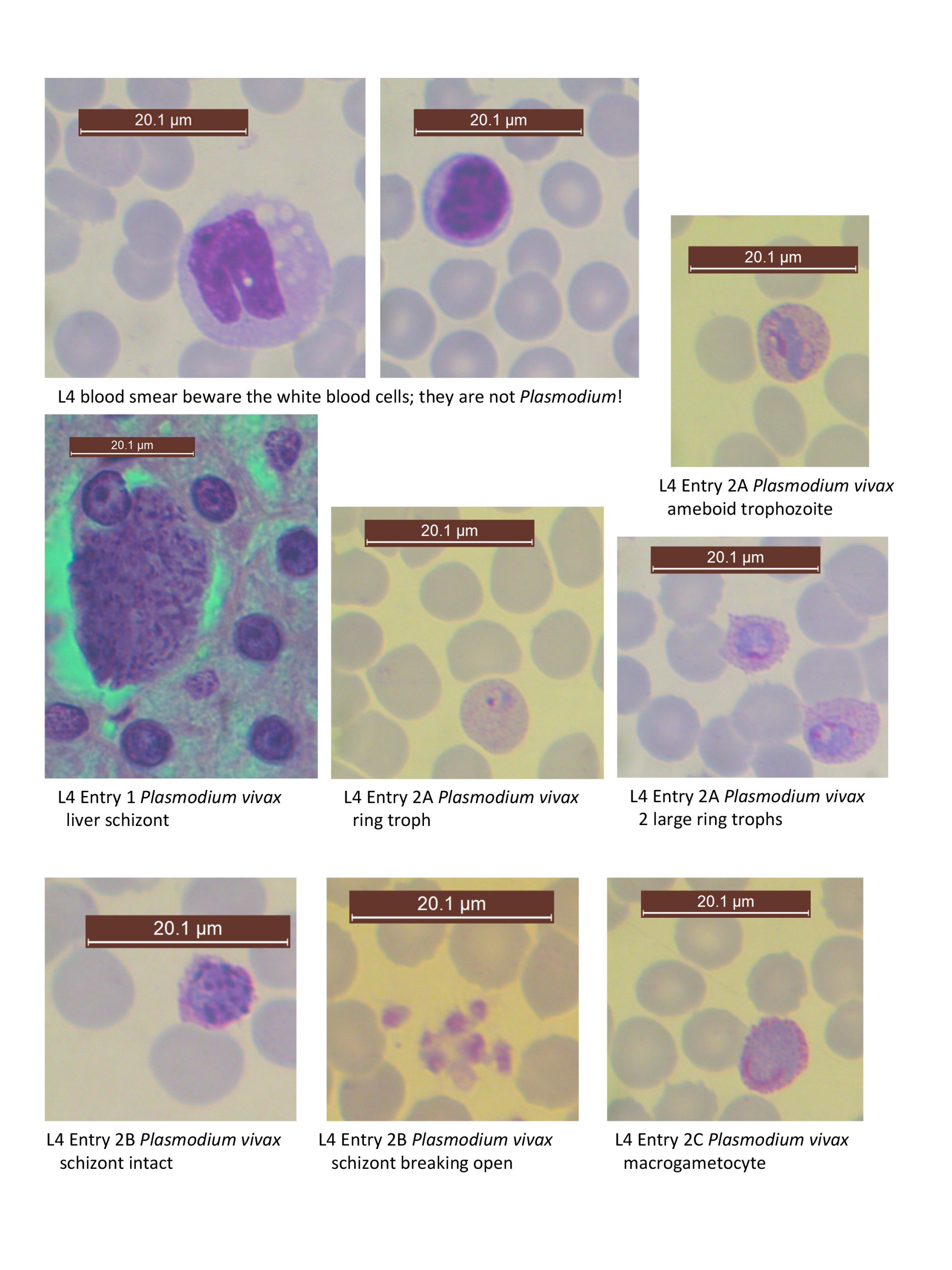
1. Plasmodium vivax preerythrocytic schizont in liver tissue. Demonstration. See text 147 Fig. 9.2. A mosquito infected with Plasmodium introduces sporozoites to the human bloodstream. After ~24 hours the sporozoites end up in the liver where they invade hepatocytes (liver cells). Within liver cells sporozoites undergo exoerythrocytic schizogony, i.e., they divide into multiple merozoites.
What ontogenetic stage came before the stage shown here in the liver? _______________
2. Plasmodium vivax. Various stages. Human blood smear. Slide box slide 10. See color plate 1. When merozoites leave liver cells they enter the bloodstream and begin the erythrocytic cycle. This begins with merozoites penetrating host erythrocytes. Upon entry, the merozoite becomes a trophozoite.
*2A. Scan the slide for trophozoites. Refer to the color plate as you search.
Does the red blood cell you located infected with a trophozoite of P. vivax contain Schuffner’s dots in its cytoplasm? ________________________
Note the characteristic “signet ring” form of this trophozoite resulting largely from the presence of a conspicuous vacuole in the center of the cell.
What do you suppose is contained in that vacuole? I.e., what is the parasite eating during this stage?_________
Be sure to illustrate both the host cell and the trophozoite. On your drawing label: red blood cell, Schuffner’s dots, and vacuole, nucleus, and cytoplasm of the trophozoite. Note the most red blood cells are only infected with a single trophozoite.
Compare infected erythrocytes with uninfected ones. Do they differ in size? __________
If so, how?____________________
*2B. Trophozoites grow and develop into schizonts with the red blood cells. If you find a good example of a schizont, please be sure to show the rest of us; they are harder to find than trophs! The schizont stage is recognized by having mutiple nuclei. You may observe dark brown pigmentation associated with the schizonts. These are hemozoin granules, waste products from the parasite’s metabolism of the hemoglobin of the host cell.
Do you see evidence of vacuoles in the schizonts?____________
*2C. Schizonts undergo schizogony resulting in multiple merozoites. Merozoites penetrate additional red blood cells and can develop as macrogametocytes or microgametocytes, or as trophozoites. Search for either type of gametocyte on your slide. The macrogametocyte fills most of the cytoplasm of the red blood cell and is generally round or oval. Microgametocytes are usually less common than macrogametocytes and are stained a lighter color. Look for Schuffner’s dots, enlargement of infected red blood cells, and look for the nucleus, cytoplasm and hemozoin granules associated with the macrogametocyte.
Find a gametocyte and describe how you would distinguish it from the other stages:______________
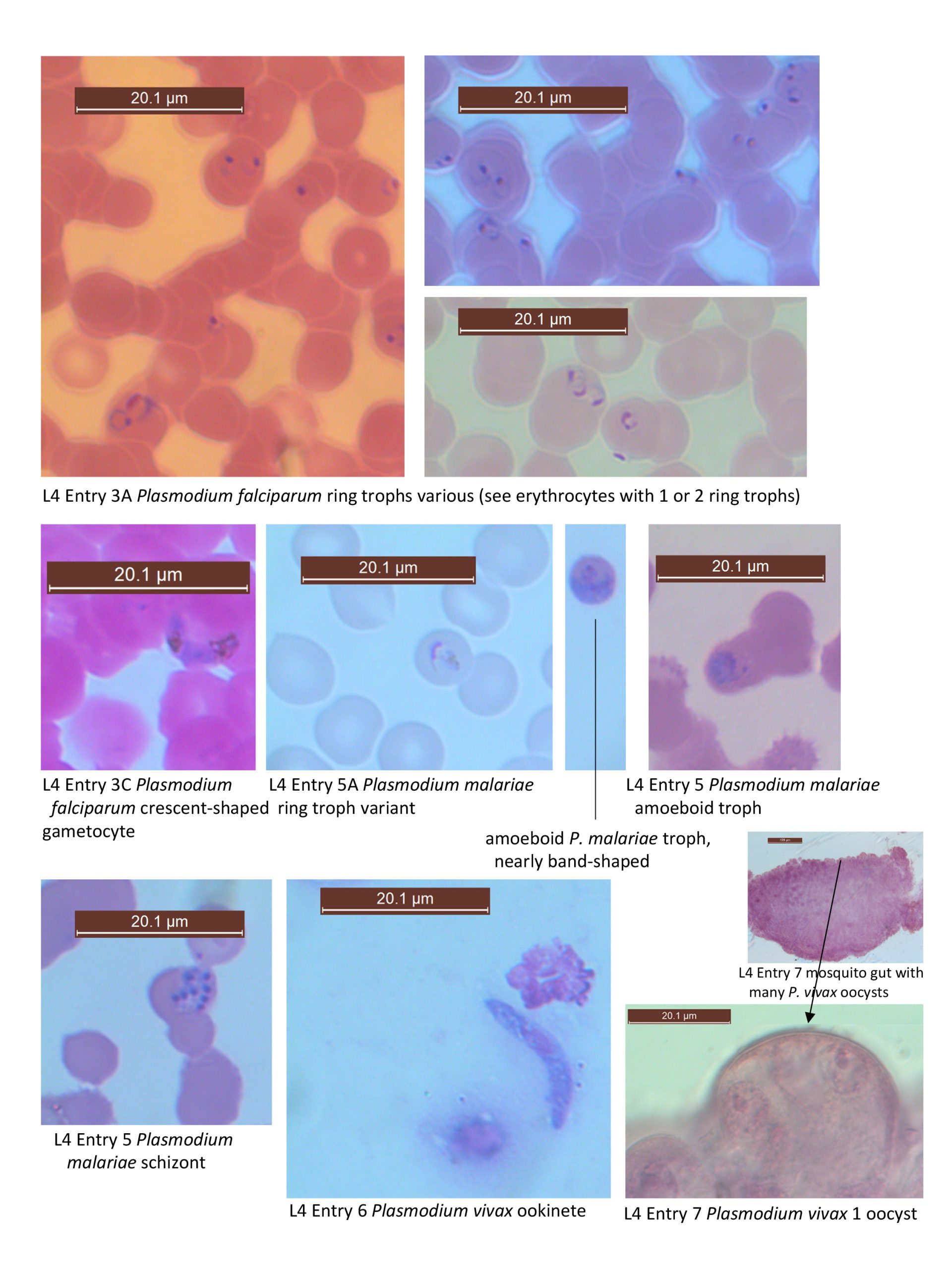
3. Plasmodium falciparum. Various stages. Human blood smear. Slide box slide 11 See color plate 3.
*3A. Plasmodium falciparum can be distinguished from other species of Plasmodium based on the relatively small size of its ring trophs and the frequent occurrence of multiple trophozoites per red blood cell. Scan your slide for trophozoites.
Do you see evidence of Maurer’s clefts? __________Compare infected blood cells to uninfected ones; do the infected red blood cells look enlarged? ________Note the “signet ring” form of the trophs, a result of the presence of a conspicuous vacuole in the center of the cell.
3B. You may also encounter schizonts of P. falciparum, but these are generally rare in peripheral blood.
3C. Look for either macrogametocytes or microgametocytes of P. falciparum. (Or see demonstration or example from another student) Both of these stages have a characteristic banana-shape, but macrogametocytes tend to be more darkly stained.
Do you see evidence of Schuffner’s dots or hemozoin?______________
What ontogenetic stage follows the macro- or microgametocyte stage?______________
4. Plasmodium falciparum in brain tissue. Histological section. Demonstration. See text Fig. 9.7. This condition results in cerebral malaria, in which capillaries are filled with red blood cells infected with Plasmodium falciparum.
What is the source of pigment that is visible in this slide?________________
5. Plasmodium malariae. Various stages. Human blood smear. Slide box slide 12
See color plate 5.
*5A. Search for trophozoites of P. malariae. These closely resemble trophozoites of P. vivax. However, some P. malariae trophozoites are band-shaped, rather than ring-shaped. The trophozoites of P. vivax are never band-shaped.
Do you see evidence of Schuffner’s dots or enlargement of infected red blood cells?________________
II. Plasmodium spp. Life cycle stages in the invertebrate host
6. Plasmodium vivax ookinetes. Demonstration slide- smear taken from the stomach of a mosquito. Once ingested by the mosquito, a single macrogametocyte develops into a single macrogamete. However, a single microgametocyte will undergo a process known as exflagellation and form 6-12 microgametes. Microgametes swim around the mosquito gut until they find a macrogamete, which it penetrates and fertilizes. The product of that union is a zygote that is elongate in form, known as an ookinete.
Are ookinetes haploid or diploid?____________________________________________
Based on the life cycle, would you consider the mosquito host the intermediate or definitive host? Explain your answer:_________________________________________
_______________________________________________________________________
7. Plasmodium vivax oocyst. Demonstration slide- sections of mosquito stomach infected with multiple oocysts. See text page 148 Fig. 9.3. The ookinete moves to the mosquito stomach wall, penetrates cells, and transforms into an oocyst. The first division within the oocyst is reductional, followed by repeated mitosis. After a series of divisions, the oocyst contains thousands of sporozoites.
What ontogenetic stage(s) in the Plasmodium life cycle are diploid?_________________
What type of process takes place within the oocyst (sporogony, gametogony, or schizogony)?__________________________
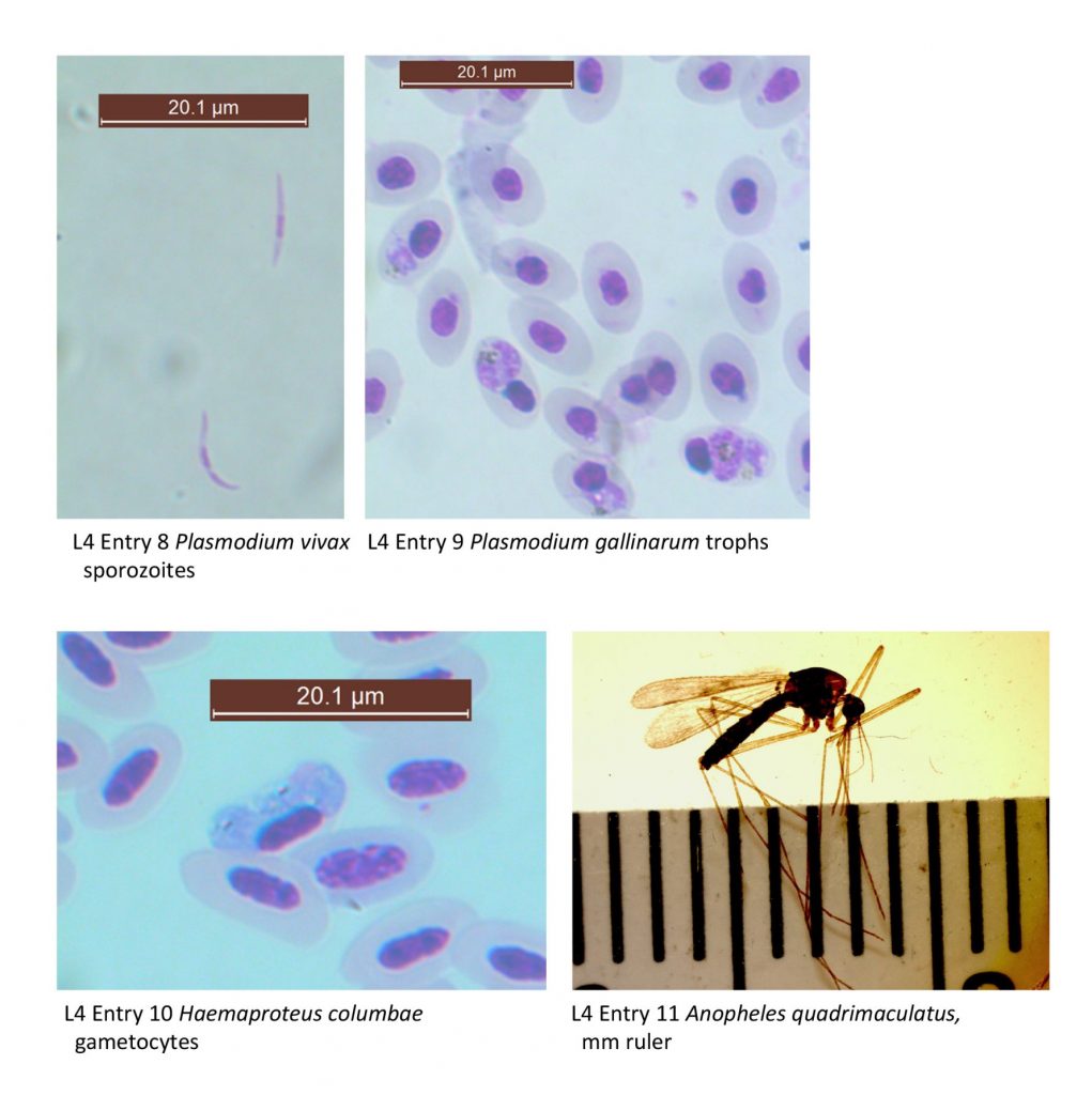
8. Plasmodium vivax sporozoite. Demonstration slide. Smear taken from the salivary gland of a mosquito. See text page 148 Fig. 9.4. Sporozoites emerge as the oocyst ruptures. They migrate from the mosquito body cavity to the salivary glands. Once a mosquito has sporozoites in its salivary glands, it is infective for the rest of its life, which is long enough to include multiple blood meals, unfortunately.
Describe the morphology of the sporozoite that you see:____________________
III. Plasmodium and related species in other animals.
Species of Plasmodium infect many other animals besides humans.
9. Plasmodium cathemerium or Plasmodium gallincaeum trophs. Bird blood smear. Slide tray. Locate the small blue bodies in the cytoplasm of the red blood cells. These small bodies are trophs of P. cathemerium. Also, try to locate schizonts.
10. Haemoproteus columbae gametocyte. Pigeon blood smear. Demonstration. See text page 159 Fig. 9.11. Species of this genus can infect a variety of birds. Note how the gametocyte occupies the majority of the cytoplasm.
Is this stage infective to other pigeons? ____________________________________
What is the infective stage of H. columbae to the pigeon?______________________
IV. An invertebrate (or “definitive”) host of species of Plasmodium
11. Anopheles quadrimaculatus. Whole mount. Demonstration. See page 582 Fig. 39.6. This is one of many species in the genus that Plasmodium species can infect. When this female mosquito takes a human bloodmeal containing gametocytes of Plasmodium, it becomes infected and the cycle continues in its gut. Non-suitable mosquito hosts would digest gametocytes along with the rest of the blood meal at this point.
List all of the ontogentic stages of the Plasmodium life cycle that occur within the lumen of the mosquito host gut/intestine:________________________________________ and,
list all of the stages that occur in other mosquito body parts:_______________________