Laboratory #5: Apicomplexa 2
Phylum Apicomplexa (Part II)
In the first part of today’s lab we will examine a species from another order in the Class Aconoidasida. The classification is shown here, with respect to the apicomplexans we saw last week:
Phylum Apicomplexa – possess an apical complex
Class Aconoidasida – no syzygy; intracellular reproduction
Order Haemospororida –heteroxenous; naked sporozoites
Plasmodium spp., Leucocytozoon sp., Hemoproteus sp.
Order Piroplasmorida – Members of this order are parasites of ticks and mammals. Trophozoites dwell within the mammal host erythrocytes. Unlike Plasmodium, there is no exoerythrocytic phase in the mammal host (e.g., see text page 163 Fig. 9.16). This order is of considerable veterinary importance.
1. Babesia bigemina. Blood smear. Demonstration of trophozoites (= “merozoites”). Text page 162 Fig. 9.15. This species parasitizes cattle, or other ruminants, and tick species of Boophilus. It is the etiological agent of a disease in cattle called “Texas cattle fever” or “red-water fever”, which can result in death within a week of infection. Observe the merozoites, which are often found in pairs in red blood cells. Hence the species name.
Refer to the life cycle of another species of Babesia, shown on text page 163:
What stage is infective to the tick, from the mammal host?______________________
What stage is infective to the mammal host?__________________
Class Conoidasida
Unlike the previous apicomplexan class you studied, members of this group possess an apical complex with a conoid.
Subclass Coccidiasina – coccidians. These parasites typically occupy sites other than blood in their vertebrate hosts, and lack syzygy. Sporozoites are usually enclosed in sporocysts within an oocyst. Some members of this group are monoxenous while others are heteroxenous.
Oocyst morphology is important for species identification. For example, Eimeria species all possess sporulated oocysts that contain 4 sporocysts, each with 2 sporozoites. Whereas the sporulated oocysts of members of the genera Toxoplasma and Sarcocystis contain 2 sporocyts, each with 4 sporozoites.
Eimeria is monoxenous, while Toxoplasma and Sarcocystis are heteroxenous.
2. Eimeria stiedae in rabbit liver. Various life cycle stages. Demonstration. Text page 129 Fig. 8.9 & attached Fig. 1. This is very similar to the species Eimeria tenella, which infects chickens and causes a high level of mortality in young birds. Eimeria stiedae is also harmful and can cause lesions in the rabbit host liver.
It is possible to observe the following stages in the liver section: schizont, macrogametocyte or microgametocyte, and unsporulated oocyst. Of these, which do you see in this field of view (list them)?____________________________________.
Answer the following two questions based on what you know about the life cycles of Eimeria species,:
Which stages of E. stiedae could occur inside liver epithelial cells?__________________
Which stages of E. stiedae could occur on the outside of the cells (i.e., free within the bile duct)?______________________
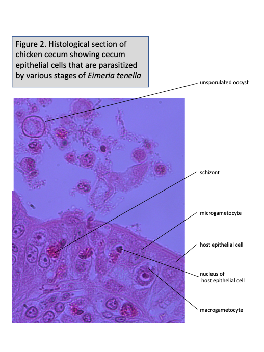
3. Eimeria tenella. Section through chicken cecum with various life cycle stages. Slide box slide 13. Text page 129 & attached Figs. 1 and 2. You should be able to locate each of the following stages. Note that you may draw these all together on a single page:
*3A. Schizont. Find a schizont within an epithelial cell of the cecum section you are viewing. Identify the merozoites within the schizont as well as the epithelial cell and epithelial cell nucleus.
*3B. Macrogametocyte. Locate a mature macrogametocyte within an epithelial cell. Identify the macrogametocyte, peripheral bodies, epithelial cell, and epithelial cell nucleus.
*3C. Microgametocyte. Locate a mature microgametocyte within an epithelial cell. Identify the microgametocyte, epithelial cell, and epithelial cell nucleus.
*3D. Unsporulated oocyst. Find a mature, unsporulated oocyst. Be sure to identify the oocyst and oocyst wall. Note that some oocysts are within epithelial cells, while some are free beyond the epithelial cells of the section you are viewing. Each oocyst breaks free from its epithelial cell, enters the lumen of the cecum and eventually pass to the outside via the feces of the host. Why wouldn’t you expect to find sporulated oocysts on the slide of E. tenella in a chicken cecum?_________________________________
4. Toxoplasma gondii. Tachyzoites. Demonstration. See text page 134 Fig. 8.14 for examples of tachyzoites, and text page 133 for the life cycle. Tachyzoites are small, merozoite-like stages. Once known only from tissue stages in the brain and other organs, Toxoplasma was shown in 1970 to undergo typical coccidian merogony (=schizogony) and gametogony in the intestine of cats, and to produce oocysts in the feces. This demonstration slide is a tissue smear from the acute phase of an infection. Note the small size of these organisms.
*5. Sarcocystis tenella. Zoitocysts in sheep muscle. Slide box slide 14. See text page 138 Fig. 8.18 for the life-cycle of another species of Sarcocystis. The life cycles of species of Sarcocystis are now known to include 2 vertebrates: a herbivore and a carnivore. These slides show zoitocysts typically known as “rice grain” cysts from the intermediate host. Note that this stage is large enough to see with the unaided eye, unlike other protist stages you have seen in lab to date! It should be drawn at your lowest magnification. The life cycle of members of this genus was not completely known until 1972.
What life cycle stage is found inside these zoitocysts?___________________________
What sort of animal might ingest a zoitocyst?_______________________________
6. Cryptosporidium parvum. Smear. Demonstration. Text page 123 Fig. 8.4. Species in this genus parasitize the brush border of the epithelial cells of the gut. Note the tiny size of these organisms. This species infects humans, and is especially problematic in immune-compromised individuals, and in children. This slide contains oocysts.
Subclass Gregarinasina – gregarines. The mature gamonts (gametocytes) of this group dwell within extra-cellular sites within their hosts. They are generally large and most possess a conoid modified into a mucron or epimerite for attachment to host cells. Schizogony does not take place in the gregarine life cycle. Gregarines do typically undergo syzygy, or the pairing of gametocytes, during the course of sexual reproduction. The paired gametocytes of many gregarines are surrounded by a protective gametocyst. All species are monoxenous parasites of invertebrates, typically occupying the digestive tract, body cavity or seminal vesicles.
We should have an opportunity to observe living gregarines later in the course. There are potentially many undiscovered species of gregarines, even locally!
7. Monocystis lumbricus. Oocysts or spores. Demonstration slide-smear from earthworm seminal vesicle. See text page 121 Fig. 8.2 for the life cycle. This is an example of an acephaline gregarine. Its body consists of a single unit. This slide contains a gametocyst containing many oocysts, each of which contains 8 sporozoites.
What reproductive process is present in Eimeria but is lacking in Monocystis?
______________________________
8. Gregarina sp. Trophozoites (=trophonts). Slide box slide 15-smears from damselfly gut. See attached Fig. 3 & text page 122 Fig. 8.3. This is a cephaline gregarine. Identify the epimerite, protomerite, and the deutomerite containing the nucleus in each individual. Why do you suppose gregarines are so diverse?_________________________
Practice Quiz:
___________________
___________________
___________________
___________________
___________________
___________________
___________________
___________________
___________________
___________________