Laboratory #7: Platyhelminthes I
Phylum Platyhelminthes — flatworms
Members of this phylum are dorso-ventrally flattened. They are also bilaterally symmetrical and lack a body cavity (i.e., they are acoelomates). The majority of species lack an anus, rather, they have a blind gut and thus food does not flow uni-directionally through their body. Instead, food enters and solid wastes exit through the mouth (if present). Most species are hermaphroditic.
This phylum consists of both free-living and parasitic species. Dugesia, also known as planaria, is an example of a free-living species that is often used in introductory biology and zoology courses.
“turbellarians” Turbellarians are not a monophyletic group, rather, they represent several early branches in the evolutionary history (phylogeny) of Phylum Platyhelminthes. This assemblage consists of the mainly free-living predator (i.e., non-parasitic) species of the Phylum Platyhelminths. Some “tubellarians” live in symbiosis as commensals. Members of this assemblage lack a neodermis.
1. Temnocephala species. Adult. Demonstration. (See text pg. 197 and Fig. 13.5 on pg. 198). This specimen was collected in southeaster Peru, from the outside of an amphipod in a tributary of the Amazon river. It is a species with several tentacles around the oral end (and hence referred to by some as the “Hamburger helper worm”). This unidentified, and perhaps new, species most likely uses the crustacean simply as a substrate for attachment, and uses its mouth to feed on smaller organisms or detritus.
Subphylum Neodermata
The four major groups of platyhelminths that parasitize vertebrates (Aspidogastrea, Digenea, Monogenea, and Cestoda) form a cohesive group, which is considered more derived than free-living species of platyhelminths. These four groups are considered to be monophyletic, and are collectively known as the Subphylum Neodermata, because of their possession of a neodermis (see below).
All species of this subphylum are obligate parasites at some point in their life cycles. In early developmental stages, members of this group possess an outer body layer in the form of a normal epidermis, i.e., a single layer of ciliated cells. However, when their first host is encountered, the ciliated epidermis is shed and replaced with a syncytial layer called the neodermis. A neodermis is considered syncytial because the cytoplasm of the cells in this layer is not separated by cell membranes—rather, it is continuous throughout the entire surface of the animal. The nuclei of these cells are located below the outer muscle layers. See texbook page 309.
References:
Caira, J. N. and D. T. J. Littlewood. 2001. Worms, Platyhelminthes. Encyclopedia of
Biodiversity 5: 863-899. Academic Press.
Carranza, S., J. Baguna and M. Riutort. 1997. Are the Platyhelminthes a monophyletic
primitive group? An assessment using 18S rDNA sequences. Molecular Biology
and Evolution 14: 485-497.
Littlewood, D. T. J., K. Rohde, R. A. Bray, and E. A. Herniou. 1999. Phylogeny of the
Platyhelminthes and evolution of parasitism. Biological Journal of the Linnean
Society 68: 257-287.
Rieger, R. M. 1998. 100 years of research on “Turbellaria” Hydrobiologia 383: 1-27.
Class Trematoda
There are 2 classes of neodermatans. Members of Class Trematoda generally possess a ventral, muscular sucker, which is sometimes subdivided into loculi.
Subclass Aspidogastrea
Species in this group parasitize molluscs, or occasionally fishes or turtles. The key feature is a large, ventral adhesive disk that is subdivided by muscular septa into individual chambers, known as loculi.
2. Aspidogaster conchicola. Adult. Demonstration. From pericardial cavity of a freshwater clam (Text pg. 204 Fig. 14.6). Be sure to recognize
the massive, ventrally located adhesive disk, as well as the buccal funnel, pharynx, tubular gut, and single ovary and testis.
Does this stage of this animal possess a neodermis?______________________
Is this animal monoecious or dioecious?____________________________
Subclass Digenea
The Digenea are commonly known as flukes. They generally use two or more hosts in their life cycles. Almost all digeneans use a mollusc as their first intermediate host. Definitive hosts are generally vertebrates, but there are some exceptions in which invertebrates, such as arthropods, serve as definitive hosts.
Digeneans are characterized by their possession of a series of unique larval forms including: miracidium, sporocyst, redia, cercaria, and metacercaria. The digeneans consist of thousands of species and >2500 genera, all of which are parasitic. In the first portion of this lab you will be introduced to the various larval life cycle stages of the flukes.
1st part of lab: Larval forms
The following entries serve as examples of typical digenean life cycle stages, in order. There are, however, a number of exceptions to this generalized theme, in which certain stages have more than one generation, or, are lacking.
Do not worry about species identities for this portion of the lab; learn the stages themselves, and their features, so that you have a general understanding of each stage as part of the digenean life cycle:
*3. Egg (same as #13). Slide box slide 16. (see text 259 Fig. 17.6). This is an egg of the species Fasciola hepatica. Note the operculum at one end of the egg. Be sure to label in your drawing: egg shell, operculum, and developing miracidium.
What function does the operculum serve?____________________________
What life cycle stage came before this one?___________________
What life cycle stage follows this one?___________________
*4. Miracidium. Slide box slide 17. (see examples in Figures in text pg. 221). This is a miracidium of Fasciola hepatica. The miracidium is one of the digenean life cycle stages that are usually free swimming.
Do you see cilia? __________Eyespots? _____________An apical organ?___________. Look in the posterior half of the specimen and locate individual germ balls. What will each germ ball become (hint, think of what stage comes two stages after this one)?__________________. Does this miracidium possess a neodermis?_____________
*5. Sporocyst. Slide box slide 18. (see figure in text pg. 222). These are sporocysts of an identified species of trematode collected from the snail Physella heterostropha as part of a student research project in my lab. Sporocysts are essentially sacs that contain many individuals of the next generation within.
What stage preceded (came before) this one?_________________
Examine the sporocysts with the microscope. What stage is visible inside each sporocyst?__________________________
*6. Redia. Slide box slide 19. (see figure in text pg. 222). These are redia of Cryptocotyle lingua. Note the mouth and obvious pharynx in this life cycle stage. Like sporocysts, redia are essentially sacs that contain many individuals of the next life cycle stage developing within.
What life cycle stage is developing within these redia?_______________________
In what kind of host animal would you seek redia?__________________________
*7. Cercaria. Slide box slide 20. (see text pg. 224 Fig. 15.24m). Cercaria are diverse in form and are therefore classified in terms of their body types. Examples of cercarial types are shown in Fig. 15.24 in your text. This is a cercaria of Schistosoma mansoni. Schistosome cercaria are termed apharyngeate furcocercous cercaria, i.e., they lack a pharynx, and have a forked tail.
Based on the morphology of this cercarial type, how do you think it encounters the next host in its life cycle (crawling or swimming)?_________________________________
*8. Metacercaria. Slide box slide 21. This is a metacercaria of Clinostomum marginatum (or “Yellow grub”, see figure) from muscle of a Pumpkinseed sunfish. This is a metacercaria that is commonly found in local fish of economic importance (e.g., Largemouth bass). Metacercaria of C. marginatum can encyst but they are commonly found freely creeping about the body cavity of the fish, or in the muscle, or on the fins. Metacercaria of other digenean species are nearly always enclosed in a bag-like cyst (see attached Fig. 5). In any case, these specimens were mounted after having been removed from the cyst. For this species, fish are considered the second intermediate host. Herons serve as the definitive hosts, while certain snail species serve as the first intermediate host. This is essentially a juvenile adult, differing from the adult only in that its reproductive system is undeveloped.
How might metacercaria differ anatomically from adult specimens of Clinostomum marginatum?______________________________________________
Examine the figure of Yellow grub, and the other metacercaria shown in the figure. Is your specimens of Yellow grub an encysted, or unencysted, metacercaria?____________
2nd part of lab: Liver flukes
In the remainder of this lab, and in lab next week, you will become familiar with adult specimens of some of the digenean species of veterinary or medical significance.
You need to be able to distinguish the adult specimens to species, as well as understand and recognize the major components of their morphology. Study the morphology of each adult example. Note that overall size and shape of the worm can be helpful. Bear in mind that many of the species of this highly diverse group are parasites of other animals, including local wildlife. Also remember that every species we discuss utilizes a snail as the first intermediate host.
The following group of digenean species you will examine in this lab are usually found associated with the liver (or gall bladder) of their definitive hosts. You should be able to identify each of these species by the end of the lab. The families to which these species belong are listed, but you are not required to learn them.
Family Opisthorchidae
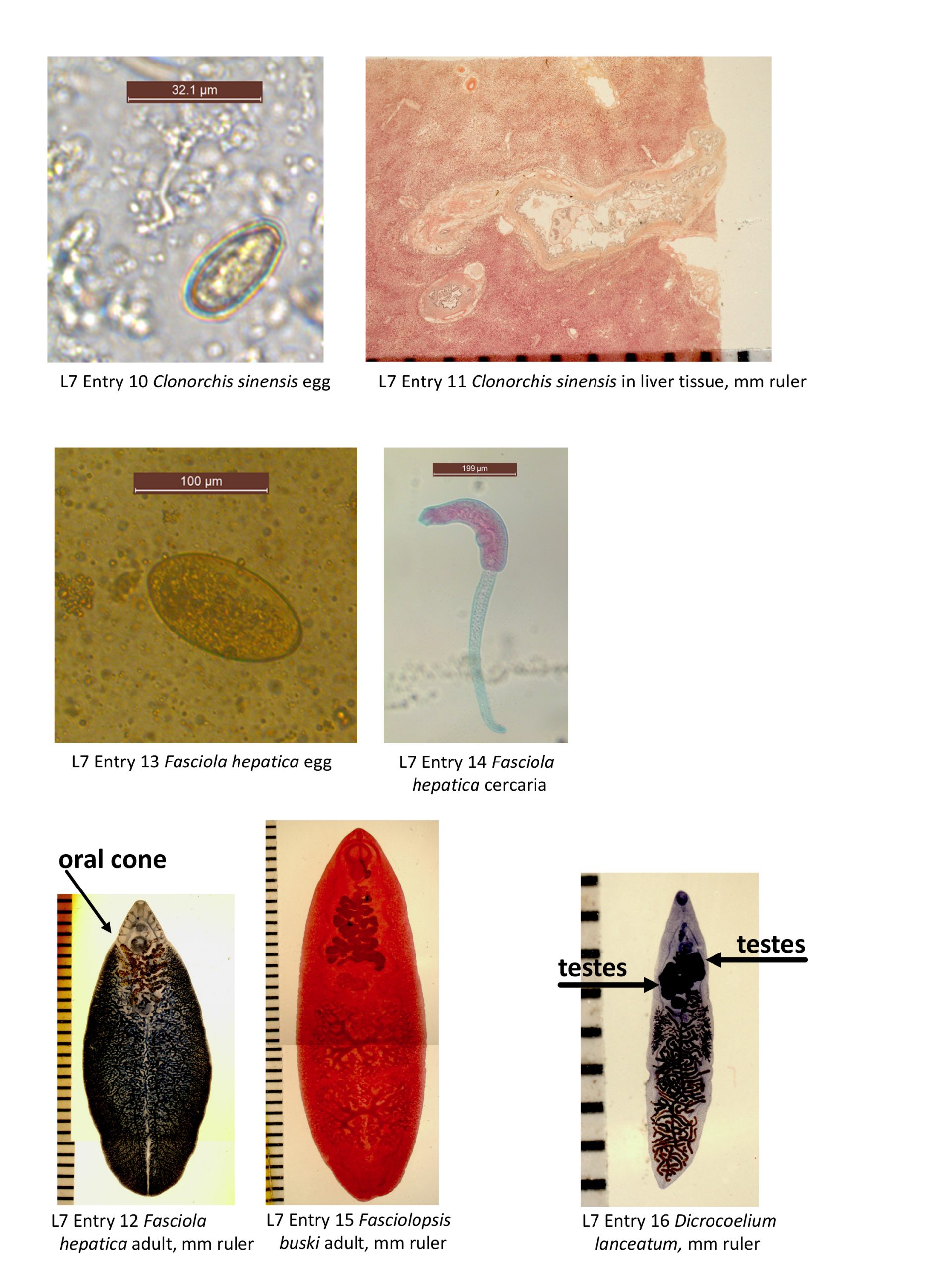
*9. Clonorchis sinensis adult. Slide box slide 22. Note that this species is referred to as Opisthorchis sinensis in some texts. (see text Fig. 18.18 pg. 276). This species is commonly known as the Chinese liver fluke. It is the etiological agent of “Clonorchiasis.”
Identification of digenean species rely on recognition of the morphological features of the adult worms, thus it is important that you understand the various structures of a typical digenean. See the morphology section of this species in your texbook for a helpful overview.
Make a full-page illustration of the adult of C. sinensis, and label the following structures: oral sucker, acetabulum (=ventral sucker), pharynx, esophagus, intestine, testes (there are 2, and are dendritic, or branched), seminal vesicle (hard to see and not required to draw), genital pore, uterus, eggs, ovary, seminal receptacle, vitelline glands, and Laurer’s canal. This plate will serve as a reference for examination of other species of digeneans.
What is the function of the vitellaria?_________________________________________
Is this specimen monoecious or dioecious?_____________________________________
How does this specimen void undigested matter?_____________________________
Does this specimen possess a neodermis?_________________________________
Does this specimen possess cilia on its outside surface?__________________
10. Clonorchis sinensis eggs. Demonstration. (see text pg. 278 figure 18.20). Eggs of this species are distinguished based on their small size, and the presence of a small knob at the abopercular (i.e., end opposite the operculum)
How many microns long is the egg you viewed?_________________________________
11. Clonorchis sinensis section of adult worms in liver tissue. Demonstration. (see text figs. on pg. 279). Note the tissue reaction associated with this parasite.
What do you suppose this specimen eats within its specific habitat?__________________
Family Fasciolidae
12. Fasciola hepatica adult. (Slide box slide 23). (see text pg. 256) Please view with a dissecting microscope AND BE CAREFUL WITH THIS VALUABLE SLIDE! This is the “sheep liver fluke.” These specimens were collected from the liver of either a sheep or a cow. This species is the etiological agent of “Fascioliasis”, which can result in gross pathology of the liver. Note the extensively branched ceca of this species. The two testes, arranged in tandem, are also branched. Also note the oral cone giving the animal an appearance of possessing “shoulders” and the anterior position of the acetabulum (ventral sucker).
Do you see evidence of spines on the surface of this species?__________Can you distinguish the two lateral fields of follicular vitellaria from the testes?_______________ How do adults get to the liver?______________How do adults of this species differ from those of Clinorchis sinensis?_____________________
*13. Fasciola hepatica eggs. (Slide box slide 16 in boxes 1-14). (see text pg. 259) View an example of an egg of F. hepatica. Note the operculum and lack of a conspicuous abopercular knob or thickening in this species.
How does this egg differ from that of Clonorchis sinensis?______________________
How many microns long is the egg you viewed?_____
14. Fasciola hepatica cercaria. Demonstration. (see text pg. 257). This cercaria drops its tail and encysts on aquatic vegetation if encountered. If not, the cercaria merely encysts in the water. The definitive host is infected either by eating aquatic vegetation, or by drinking water containing metacercaria.
How does this aspect of the life cycle of this species differ from the life cycle of the oriental liver fluke, C. sinensis? (Hint compare text Figs. 17.5 and18.19):_____________
15. Fasciolopsis buski adult. Demonstration. (see text pg. 260). Although this species is closely related to Fasciola hepatica, it is NOT a liver fluke, rather it inhabits the small intestine of its definitive host. Note that the ventral sucker is larger than the oral sucker. Also note the unbranched ceca and dendritic (branched) testes in the posterior half of the worm.
How does this species differ from morphologically from F. hepatica? ___________
What is the definitive host of this species?_________________________________
Family Dicrocoelidae
16. Dicrocoelium dendriticum or (D. lanceatum) adult. Demonstration. (see text pg. 266). Compare the morphology of this species with that of other adult worms you have examined, and in particular with C. sinensis to which it is most similar in terms of size and shape. The natural definitive hosts of this species include sheep, goats, cattle, pigs, deer, marmots and rabbits.
In what kind of first intermediate host would you expect to find this fluke? ___________
Do the cercaria of this species actively burrow out of the first intermediate host? ____
How does the life cycle of this species exemplify a parasite’s ability to alter the behavior of its host to the parasite’s advantage?_______________________________________