Laboratory #9: Platyhelminthes III (Monogeneans and cestodes)
Phylum Platyhelminthes — flatworms
Subphylum Neodermata
Class Trematoda
Class Cercomeromorpha
Subclass Monogenea
Subclass Cestoda
Class Cercomeromorpha
Species belonging to this class possess an extension of the body known as the “cercomer”. The cercomer typically bears 3 or more pairs of hooks. This feature is present in both larval and adult stages of monogeneans, but only in larval cestodes.
I. Subclass Monogenea
Species of this group are typically ectoparasitic, and have monoxenous life cycles. They are typically found parasitizing the skin and gills of fishes. A few species parasitize amphibians and reptiles, and one species, Oculatrema hippopotami, is a parasite on the eyes of hippopotamuses. The monogeneans have a cercomer (as a haptor) in the adult stage, and eyespots in the larval stage, known as the oncomiracidium.
1. Actinocleidus sp. whole mount. Demonstration. See textbook page 292. This serves as an example of a locally collected monogenean species. This specimen was obtained from the gills of a Smallmouth bass in Otsego Lake, New York. Compare this specimen to Figure 2.
What part of the fish do you suppose serve as attachment points for the hooks that are visible in the haptor of this specimen?________________________________________
How many hosts are in the life cycle of Actinocleidus sp.?_________________________
2. Microcotyle sp. whole mount. Slide tray. MUSEUM SPECIMENS: TREAT WITH A HIGH LEVEL OF RESPECT AND CAUTION! This worm was collected from the gills or skin of a Quillback rockfish (Sebastes maliger), from the North Atlantic coast. This monogenean species belongs to a group within the Subclass Monogenea known as the Polyopisthocotylea. Polyopisthocotyleans possess a haptor that is subdivided into one or more pairs of suckers, and in this example, sclerotized attachment structures called clamps. Label the following in your drawing: haptor, clamps, testes, vitellaria, attachment organ, pharynx, gut, ovary, egg capsule, and vitelline duct.
II. Subclass Cestoda
Cestodes, or tapeworms, are endoparasites that typically occur in the gut (i.e., small intestine) of vertebrates during the adult stage. Cestodes have been reported from all classes of vertebrates but are most in common in fishes, birds, and mammals. Perhaps the most striking morphological feature of cestodes is the scolex, a holdfast organ at the anterior portion of the body. The scolex is highly variable among cestode species, and can include specialized suckers, and hooks in many cases. The cestode body consists of a chain of segments or proglottids, which each contain a complete set of male and female reproductive organs. Most cestodes produce proglottids near the posterior margin of the scolex, in a region often called the neck. The proglottids nearest the neck are immature (newest formed), and the proglottids mature as they are pushed away from the neck by the more newly formed proglottids. Mature proglottids (i.e., proglottids containing fully formed and functioning reproductive organs) are found towards the middle of the strobila. Gravid proglottids (proglottids filled with eggs) are found towards the posterior end of the strobila in tapeworms, except in species that drop proglottids. Cestodes drop their proglottids from their strobila at different points of maturity and it is not uncommon to find large numbers of individual free proglottids in a host. In general, cestodes require at least two host in order to complete their life cycles.
Cestodes also differ from other platyhelminths in that they lack a digestive system. Instead, tapeworms absorb nutrients across their neodermis, or tegument. In cestodes, the neodermis is covered with variable structures called microtriches (see textbook page 309).
You will learn some general features of the cestode body. While the proglottids of cestodes are very similar to the bodies of digeneans (i.e., a complete set of male and female reproductive structures are present), they do differ in several ways.
3. Acanthobothrium larsoni whole mount of adult. Demonstration. See figures in this manual and research article. This specimen was collected from the spiral intestine of a stingray off the coast of the Malaysian portion of the Island of Borneo. Observe the 4 bothridia of the scolex, which have muscular edges and septa that form distinct loculi. Each bothridium bears a pair of bipronged hooks. Note the relatively small size of this species. Look at the copy of the research article included in the lab. You’ll see that in it I named this species after my college parasitology instructor, Dr. Ingemar Larson.
How many proglottids are present on this worm?________________________________
Are all the proglottids mature? How can you tell?________________________________
4. Schizocotyle acheilognathi whole mount of adult. Demonstration. This specimen was collected from a type of minnow called a golden shiner, from near Otsego Lake here in New York. This species is well-enough known to have a common name, the Asian fish tapeworm. Asian fish tapeworm has been reported from >200 species of fish hosts from around the world. It’s originally from Asia–probably Japan–but has since been introduced to other continents by way of fish farming of food fish such as grass carp. This species is considered an invasive species because of its ability to switch to other fish hosts wherever it gets introduced. It has been reported from 20 states in the USA, including New York (see copy of published paper on its report in New York).
Describe as best you can the morphology of the scolex of this specimen:___________________________________________________________
5. Diphyllobothrium latum whole mount of gravid proglottid of adult. Slide box slide # 25. See text Fig. 20.17 pg. 310 and Fig. 21.3 pg. 326. This species is commonly known as the broad fish tapeworm. The definitive host is usually a fish-eating carnivore, such as a polar bear or human. Note the lateral follicular vitellaria. In this species, eggs are released from the uterine pore of gravid proglottids while still attached to the stobila. Examine a gravid proglottid and locate the uterine pore.
How would you distinguish this proglottid from a proglottid of T. pisiformis?_________________________________________________________
6. Diphyllobothrium latum whole mount of eggs. Demonstration. (See text pg. 326 Fig. 21.4). Study the representative egg. Recognize the following structures: hexacanth, shell, operculum, abopercular knob. The mature oncosphere of Diphyllobothrium possesses a ciliated layer and is thus called a coracidium larva.
Is this egg ready to “hatch” in the water? ________________________________
Why or why not?________________________________________________
How would you distinguish eggs of this species from the eggs of digenean species you already examined?_______________________________________________________
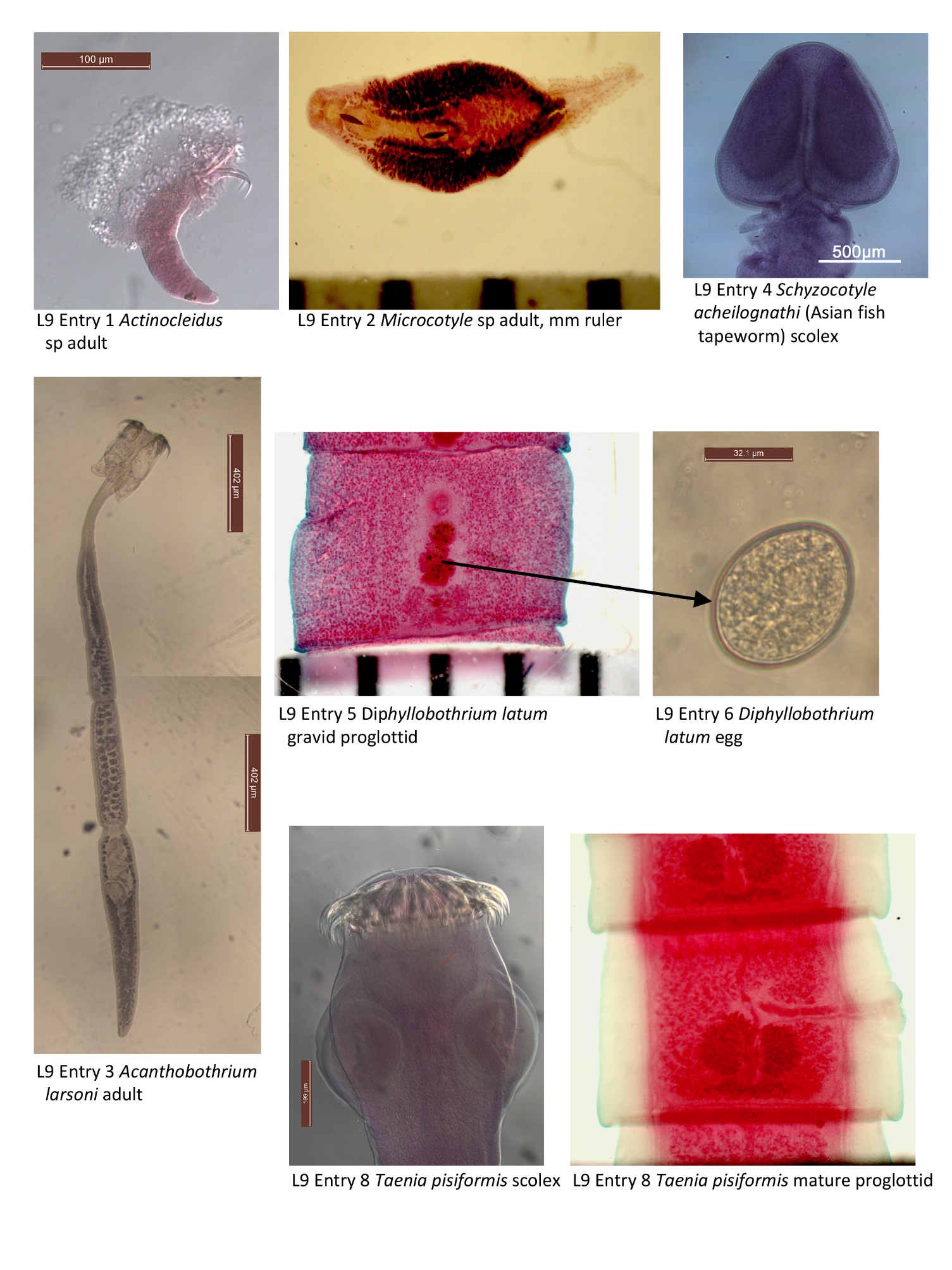
**7. Taenia pisiformis. Composite slide of adult worm. Slide box slide #26. See textbook page 310 for diagram of mature proglottid. Adults of T. pisiformis occur primarily in dogs. Dogs become infected with T. pisiformis when they consume cottontail rabbits that are infected with the larval stage of the worm. Examine the scolex with its 4 suckers and rostellum bearing numerous hooks (see scolex of similar species on text pg. 333). Make an illustration of the scolex and label hooks and suckers. Adult worms consist of 100s of proglottids and can be quite long, too long to fit on a single slide. This slide therefore includes representative immature, mature, and gravid proglottids. Try and find each of the structures labeled in attached Figure 3, and make a full-page plate of a mature proglottid—check to be certain you have chosen a mature proglottid before you invest time drawing! Also compare the uterus in mature and gravid proglottids.
What is the function of the uterus?________________________________________________________
What is the function of the vitellarium?______________________________________
8. Taenia pisiformis. Composite slide of various portions of strobili & intact worm in dish. Demonstration. This partial worm was obtained from the vomit of “Popcorn”, a domestic cat in Otego, New York. Observe the specimen with the dissecting microscope, as well as without the aid of a microscope, to gain a sense of its actual size. How do you suppose “Popcorn” acquired infection with this specimen?______________________________________
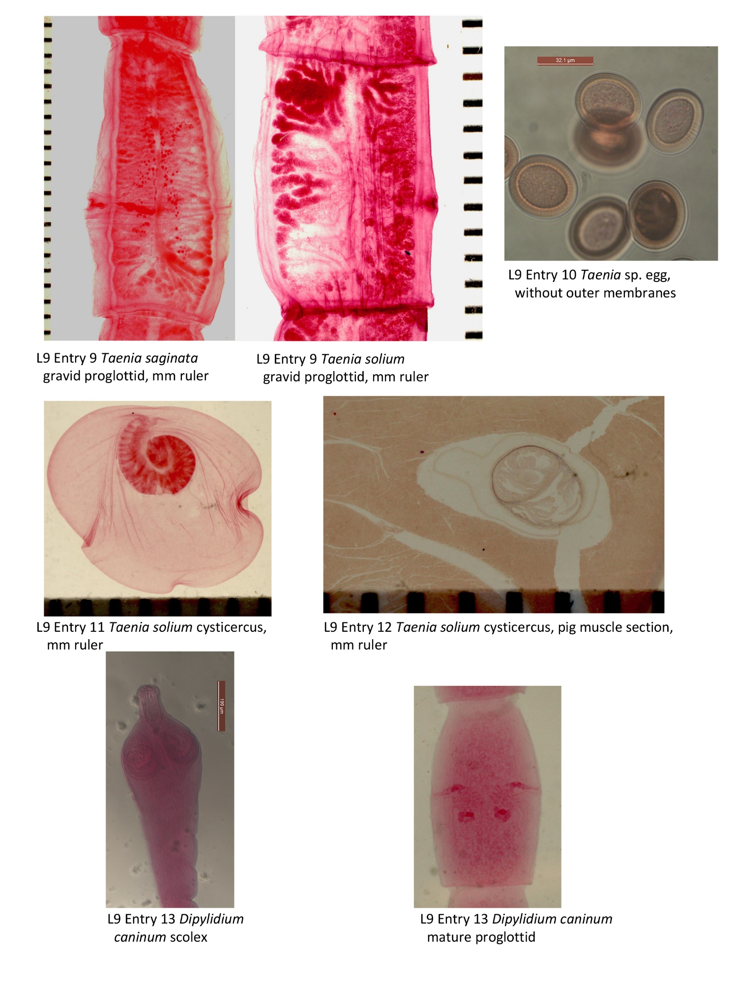
9. Taenia solium and Taenia saginata gravid proglottids. Demonstration. (See life cycle text pg. 334). Both of these species parasitize humans and possess eggs that are nearly identical. Both of these species also release detached gravid proglottids into the feces of their hosts. Thus, it is common to find gravid proglottids, rather than eggs, in the feces of individuals infected with either species of tapeworm. One of the most important features for distinguishing between infections of these two species is the number of uterine branches in the gravid proglottid. Compare the gravid proglottids of each of the two species.
Count the number of branches on the gravid uterus and specify below:
Taenia solium has _______ uterine branches.
Taenia saginata has _______ uterine branches.
10. Taenia solium or T. saginata egg. Demonstration. (See text pg. 331). This slide demonstrates an eff of a species of Taenia in which the outer envelope has been lost. While eggs of genus Taenia have a distinctive appearance, it is very difficult to distinguish between Taenia solium and Taenia saginata based only on the egg. This is why it’s very important to use gravid proglottids to distinguish between these 2 species.
Imagine you are a clinician examining a patient who has either Taenia solium or Taenia saginata. Why would you want to be sure to correctly identify the Taenia to species?_________________________________________________________
11. Taenia solium cysticercus. Whole mount. Demonstration. (See text figs. on pgs. 314 & 335). Humans can also host this ontogenetic stage by incidental consumption of eggs of T. solium.
What ontogenetic stage precedes this one in the life cycle of T. solium?____________
What ontogenetic stage follows this one in the life cycle of T. solium?____________
12. Taenia solium cysticercus-section in pig muscle. Demonstration. (See text figs. on pgs. 314 & 335). Cysticerci of this species are normally found in pork and are often referred to as “Cysticercus cellulosae”. Humans who ingest this larval stage will serve as definitive host to the adult stage of this worm.
What practice, with regards to pork, would minimize risk of infection?______________
13. Dipylidium caninum adult. Composite slide of adult worm. Demonstration. (see fig. in text pg. 343). Observe the proglottids, which are diagnostic in that they each possess two sets of male and female reproductive organs and two genital pores. The adults of D. caninum are common parasites of domestic dogs and cats and have occasionally been found in children. The larvae are simple cysticercoids that develop in fleas.
How do you suppose dogs become infected with D. caninum?_____________________________________________________________
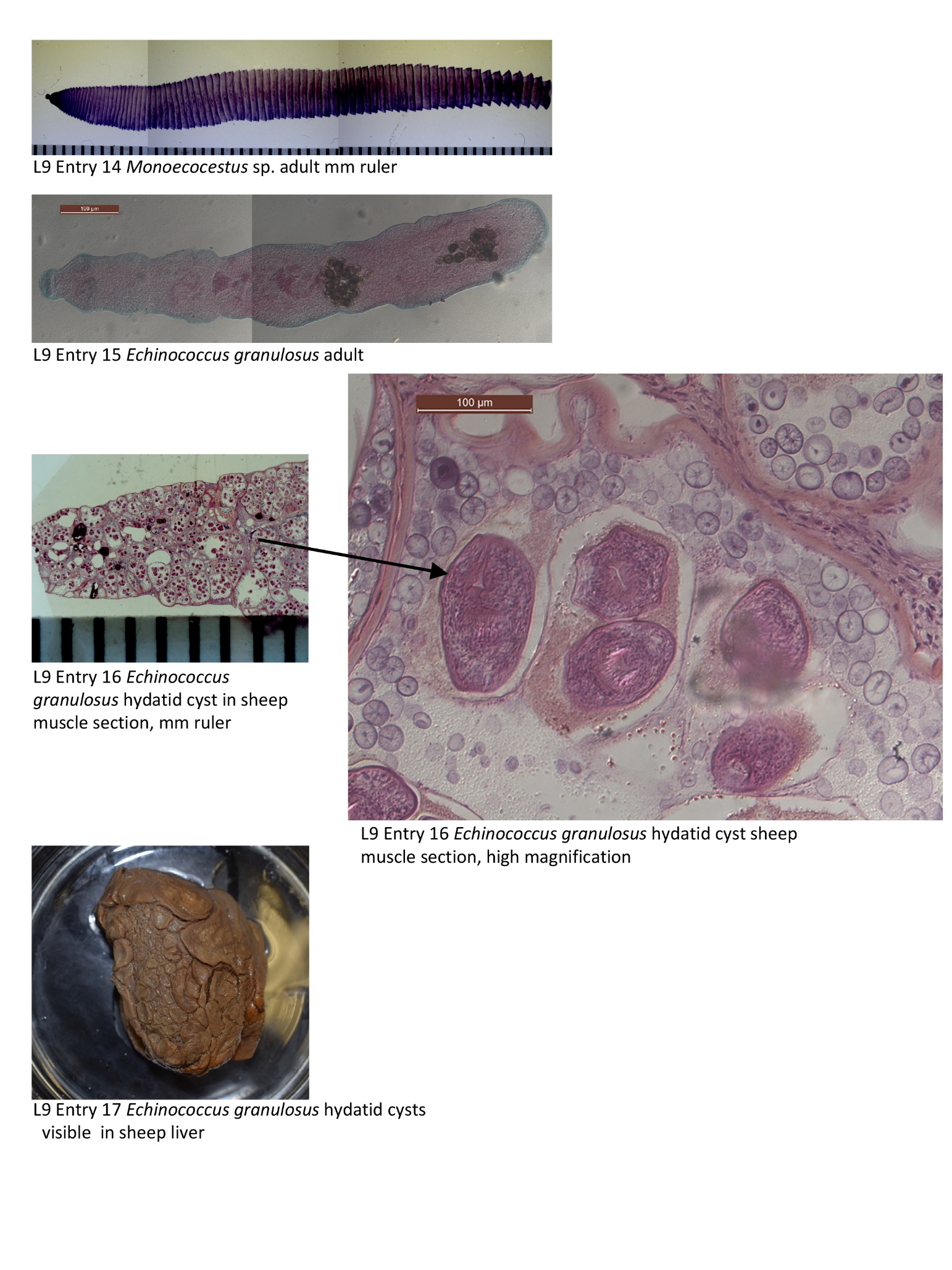
14. Monoecocestus sp. from area porcupine. Demonstration. The cestode specimens in the bottle and on the slide were obtained from 2 roadkill porcupines from Otsego County, NY, near Oneonta. Both had dozens, if not hundreds of these cestodes in the small intestine. The intermediate host is a species of orobatid mite, a tiny free-living mite that lives in the grass. Mites become infected by ingesting eggs left in the grass when the porcupine defecates. This likely happens in summer, when ground vegetation is the predominant part of the porcupine diet. Porcupines are arboreal at other times.
How do you suppose porcupines become infected with these cestodes?_______________________________________________________
*15. Echinococcus granulosus adult from intestine of dog or other carnivore. Whole mount. Slide box slide #27. (See text pgs. 337-339). Adult individuals of this species are very small, consisting solely of a scolex, a short neck, and a total of only three proglottids. Adults are parasites of the intestinal tract of canids and felids.
How many proglottids are present in this specimen?______________________________
Does the scolex of this specimen more closely resemble Acanthobothrium larsoni, or Taenia pisiformis?_____________________________________
16. Echinococcus granulosus histological section of sheep muscle showing wall of unilocular hydatid cyst. Demonstration. See text pgs. 337-339. Larvae, in the form of unilocular hydatid cysts, are generally found in mammals such as sheep and caribou. Unilocular hydatid cysts are generally significantly larger than the adult worm; one record exists of a unilocular hydatid cyst with up to 40 liters of fluid! Humans become infected with unilocular hydatid cysts by consuming eggs of E. granulosus. The cyst consists of a fluid-filled bladder surrounded by a cyst wall that is lined with a germinative epithelium. This germinitive layer can produce several million prtoscoleces in a single common bladder. Budding within daughter cysts producing more protoscoleces is also possible. Protoscoleces that break free and sink to the bottom of a cyst are called hydatid sand. These scoleces are still infective to the intermediate or definitive host.
How does this larval stage differ from the cysticercus you saw of T. solium?_______________________________________________________________
17. Sheep liver with unilocular hydatid cysts of Echinococcus granulosus. Demonstration. This sheep liver is laden with many cysts of E. granulosus. Each of these contain thousands, or potentially millions, of protoscoleces that each could potentially develop into an adult worm, had this sheep (and its liver) been consumed by a carnivore such as a dog or a wolf. This demonstration should help you put into context your observations of #15, a section of one of these unilocular hydatid cysts. Remember that humans can serve as the intermediate host of E. granulosus. I.e., infected organs in humans, such as the liver or lungs, could take on a similar appearance as this example. Remember that humans cannot serve as the definitive host of this tapeworm species.
Based on diet, what type of animal do you think would consume the host of a unilocular hydatid cyst?____________________________________